 2026, Vol. 25


精索静脉曲张(varicocele,VC)是常见的男性生殖系统疾病,其危害主要在于使患侧睾丸体积减小和对精液质量的负向影响[1]。在青少年中VC的发病率达14% ~20%,与成人相似[2]。现有研究表明对青少年VC的治疗可以有效增加精子浓度和睾丸体积,使患侧睾丸在治疗后出现“追赶性生长”[3]。腹腔镜Palomo术被认为是治疗青少年VC的一种安全且有效的手术方法[4], 但术后鞘膜积液发生率较高,可能与术中精索淋巴管的损伤有关[5]。近年来吲哚菁绿(indocyanine green,ICG)被广泛应用于外科手术以辅助定位淋巴组织[6]。本研究通过在腹腔镜Palomo术中使用ICG辅助保留精索淋巴管,以探讨该方法的应用效果。
资料与方法 一、临床资料本研究为回顾性队列研究,纳入2025年1月至2025年7月在首都医科大学附属首都儿童医学中心泌尿外科手术治疗的VC患儿。研究对象纳入标准:①年龄9~17岁;②原发性VC并符合下列手术指征之一:术前双侧睾丸体积差>20%,术前精索静脉内径>2.7 mm(Ⅱ度、Ⅲ度),伴有阴囊坠胀或睾丸疼痛;③术前患侧无睾丸鞘膜积液;④既往患侧无腹股沟、腹膜后或睾丸手术史。排除标准:临床资料不完整。收集的临床资料包括年龄、患侧、分级、淋巴管显影情况、淋巴管条数、精索静脉内径、睾丸体积、手术相关并发症。将患儿分成ICG辅助腹腔镜保留淋巴管的Palomo术组(ICG组,n=24)和传统腹腔镜Palomo术组(对照组,control group, CG组,n=31)。进一步按术前双侧睾丸体积差将患儿分成术前双侧睾丸体积差>20%的ICG辅助腹腔镜保留淋巴管的Palomo术组(ICG-A组,n=10)和术前双侧睾丸体积差>20%的传统腹腔镜Palomo术组(CG-A组,n=11);按术前精索静脉内径将患儿分成术前精索静脉内径>2.7 mm的ICG辅助腹腔镜保留淋巴管的Palomo术组(ICG-B组,n=18)和术前精索静脉内径>2.7 mm的传统腹腔镜Palomo术组(CG-B组,n=18)。本研究获得首都医科大学附属首都儿童医学中心伦理委员会批准(SHERLL2024132)。
二、手术方法患儿均采用全身麻醉,仰卧位,常规消毒铺巾。取脐部切口约1 cm,置入5 mm Trocar,建立CO2气腹(压力10 mmHg,1 mmHg=0.133 kPa,流量4 L/min),置入腹腔镜。平脐部左右两侧与腹直肌外缘交点处各取切口约0.5 cm,在腹腔镜监视下分别穿刺置入5 mm Trocar。于患侧内环口上方2~3 cm处剪开后腹膜暴露精索血管束。ICG组:将1瓶ICG(25 mg) 用10 mL灭菌注射用水稀释成2.5 mg/mL。术者一手固定患侧睾丸,一手使用26G针头垂直刺入睾丸实质,注射ICG 1 mL。在腹腔镜荧光模式下观察精索淋巴管显影情况。在荧光辅助下分离出精索淋巴管。使用Hem-o-lok结扎剩余精索血管束并剪断(图 1)。CG组:直接使用Hem-o-lok结扎精索血管束并剪断。排出腹内残余CO2,去除Trocar,缝合腹部切口。

|
图 1 睾丸内注射吲哚菁绿后淋巴管显影和术后淋巴管被完整保留 Fig.1 Lymphatic vessel visualization after intratesticular injection of indocyanine green and complete preservation of lymphatic vessels after surgery 注 A、C为白光模式;B、D为近红外光模式 |
术后记录阴囊染色情况、阴囊血肿情况和睾丸疼痛情况。术后1、3、6个月随访超声测量患侧精索静脉内径和双侧睾丸体积、查看有无睾丸鞘膜积液、睾丸萎缩和复发。精索静脉内径在患儿仰卧位静息状态下测量。睾丸体积按Lamber公式计算(长×宽×厚×0.71)。
四、统计学处理采用SPSS 27.0进行统计学分析。服从正态分布的计量资料用x±s表示,组间比较使用两独立样本t检验,组内比较采用配对t检验;不服从正态分布的计量资料用M(Q1,Q3)表示,组间比较使用Mann-Whitney U检验,组内比较采用Wilcoxon符号秩检验;计数资料用n(%)表示,组间比较使用卡方检验或Fisher精确概率法。P<0.05表示差异有统计学意义。
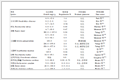
结果ICG组年龄(11.89±1.28)岁,患侧均为左侧。根据超声诊断标准[7]:Ⅰ级6例,Ⅱ级7例,Ⅲ级11例。患儿均顺利完成手术,术中至少保留1根淋巴管。术中向睾丸内注射ICG后没有患儿发生过敏反应,淋巴管均在30 s内显影,显影淋巴管数量1~4根,白光模式下呈墨绿色(图 1A、1C),近红外光模式下呈绿色荧光(图 1B、1D)。术后没有患儿出现阴囊染色、阴囊血肿和睾丸疼痛。截至术后6个月随访,ICG组无一例发生睾丸鞘膜积液、睾丸萎缩和复发。CG组年龄(11.84±1.53)岁,患侧均为左侧。Ⅰ级13例,Ⅱ级3例,Ⅲ级15例。截至术后6个月随访,7例发生睾丸鞘膜积液,没有患儿发生睾丸萎缩和复发。两组患儿年龄和分级比较,差异均无统计学意义(P=0.765,P=0.619)。ICG组术后睾丸鞘膜积液发生率明显低于CG组(P=0.015)。见表 1。
| 表 1 ICG组和CG组VC患儿一般资料和手术情况比较 Table 1 Comparison of general characteristics and surgical data between ICG and CG groups of children with VC |
|
ICG-A组和CG-A组患儿患侧睾丸体积分别在术前和术后6个月比较,差异均无统计学意义(P=0.740,P=0.852)。与术前相比,术后6个月两组患儿患侧睾丸体积均有明显增长(P<0.001)。见表 2。
| 表 2 ICG-A组和CG-A组VC患儿手术前后患侧睾丸体积比较(x±s, cm3) Table 2 Comparison of preoperative and postoperative affected-side testicular volume between ICG-A and CG-A groups of children with VC(x±s, cm3) |
|
|
ICG-B组和CG-B组患儿患侧精索静脉内径分别在术前和术后6个月比较,差异均无统计学意义(P=0.097和P=0.265)。与术前相比,术后6个月两组患儿患侧精索静脉内径均有明显缩小(P<0.001)。见表 3。
| 表 3 ICG-B组和CG-B组VC患儿手术前后精索静脉内径比较[M(Q1,Q3),mm] Table 3 Comparison of preoperative and postoperative spermatic vein internal diameter between ICG-B and CG-B groups of children with VC[M(Q1, Q3), mm] |
|
|
精索淋巴管的引流主要来源于三个部分:白膜和鞘膜脏层的浅层淋巴系统、附睾和睾丸的深层淋巴系统和部分鞘膜壁层的淋巴系统,其沿精索血管走行,最终汇入主动脉旁或主动脉前淋巴结。沿腹股沟管向上,淋巴管从7~8根减少至2~4根,这些淋巴管在腹腔内与精索血管紧密贴合直至穿过输尿管[8]。如果术中损伤这些淋巴管,会导致睾丸和鞘膜的淋巴回流受阻,造成睾丸鞘膜积液的发生。目前国内外指南均建议VC术中保留精索淋巴管[7, 9]。肉眼下的淋巴管近似无色透明,很难全部识别。为了解决这一难题,有学者提出使用亚甲蓝、异硫蓝和专利蓝等蓝色染料对精索淋巴管进行染色识别,以便辅助分离和保留淋巴管[10]。随着这些蓝色染料的使用,学者们也注意到一些与之相关的问题(如药物相关不良反应、注射部位皮肤的染色和术后尿液颜色的改变等)。淋巴管破损后局部术野的污染也是一个困扰术者的问题,一些研究更是指出注射亚甲蓝后睾丸内出现异常病变[11-12]。
ICG是一种被批准应用于临床的染料,其在特定波长的近红外光照射下可发出荧光,使用特定设备可将其接收转换成像。由于ICG几乎全部经由肝脏代谢,术后不会出现尿色的改变。目前鲜有使用ICG行淋巴造影后发生不良反应的报道。对碘过敏的人群可能是ICG唯一的使用禁忌。相比于传统蓝色染料,ICG也不存在注射部位皮肤染色和污染术野等问题。本研究中,ICG组所有患儿经睾丸注射ICG后精索淋巴管均在短时间内成功显影并顺利完成手术,没有患儿发生ICG相关药物不良反应和睾丸注射相关不良反应。与传统腹腔镜Palomo术相比,ICG辅助腹腔镜保留淋巴管的Palomo术在改善睾丸体积和精索静脉内径方面效果相当。需要注意的是,VC术后出现的睾丸“追赶性生长”可能是青春期睾丸发育的自然规律而非手术干预的直接结果。虽然本研究中ICG-A组和CG-A组术后患侧睾丸都出现了“追赶性生长”,但是由于缺少健康青少年作为对照,不能排除这一结果的出现是睾丸发育的自然规律,这也是本研究潜在的局限性。术后并发症方面,ICG组和CG组均没有患儿发生睾丸萎缩和复发,ICG组睾丸鞘膜积液发生率明显低于CG组,因此我们认为经睾丸途径注射ICG辅助保留精索淋巴管的方法是可行的。虽然使用ICG辅助可以精准定位精索淋巴管,但是在不损伤淋巴管的前提下将其与精索血管分离仍存在一定难度。我们的经验是:①对于睾丸体积较小的患儿,可根据注射ICG时睾丸内的压力适当减少注射剂量至0.5 mL,该剂量不会对淋巴管的显影产生影响;②注射ICG后可局部按压注射点30 s,以减少阴囊血肿的形成;③术中剪开后腹膜暴露精索血管后,应先使用ICG进行淋巴管造影,待淋巴管显影后再游离精索血管,以避免操作时对淋巴管的误损伤;④分离淋巴管时应操作轻柔,切勿使用电刀和钳夹,以避免损伤淋巴管;⑤术中应至少保留1根淋巴管。
综上所述,在腹腔镜Palomo术中使用ICG辅助保留精索淋巴管是安全、有效的,可以显著降低睾丸鞘膜积液的发生。该方法可以快速显影、定位精索淋巴管。相信未来随着荧光腹腔镜系统的普及,该方法在腹腔镜手术治疗青少年VC中会有更加广泛的应用。
利益冲突 所有作者声明不存在利益冲突
作者贡献声明 文献检索为徐谊、崔晓梦,论文调查设计为刘晓萌、白东升,数据收集与分析为张晨捷,论文结果撰写为刘晓萌,论文讨论分析为刘晓萌、白东升
| [1] |
Nork JJ, Berger JH, Crain DS, et al. Youth varicocele and varicocele treatment: a meta-analysis of semen outcomes[J]. Fertil Steril, 2014, 102(2): 381-387. e6. DOI:10.1016/j.fertnstert.2014.04.049 |
| [2] |
Silay MS, Hoen L, Quadackaers J, et al. Treatment of varicocele in children and adolescents: a systematic review and meta-analysis from the European association of urology/European society for paediatric urology guidelines panel[J]. Eur Urol, 2019, 75(3): 448-461. DOI:10.1016/j.eururo.2018.09.042 |
| [3] |
Locke JA, Noparast M, Afshar K. Treatment of varicocele in children and adolescents: a systematic review and meta-analysis of randomized controlled trials[J]. J Pediatr Urol, 2017, 13(5): 437-445. DOI:10.1016/j.jpurol.2017.07.008 |
| [4] |
Ulusoy O, Karakus OZ, Ateș O, et al. Successful outcomes in adolescent varicocele treatment with high-level laparoscopic varicocelectomy[J]. J Pediatr Surg, 2020, 55(8): 1610-1612. DOI:10.1016/j.jpedsurg.2019.07.008 |
| [5] |
Tandon S, Bennett D, Mark Nataraja R, et al. Outcome following the surgical management of varicocele in children and adolescents: a systematic review and meta-analysis[J]. Ther Adv Urol, 2023, 15: 17562872231206239. DOI:10.1177/17562872231206239 |
| [6] |
Zhou SC, Tian YT, Wang XW, et al. Application of indocyanine green-enhanced near-infrared fluorescence-guided imaging in laparoscopic lateral pelvic lymph node dissection for middle-low rectal cancer[J]. World J Gastroenterol, 2019, 25(31): 4502-4511. DOI:10.3748/wjg.v25.i31.4502 |
| [7] |
中华医学会男科学分会精索静脉曲张诊断与治疗指南编写组. 精索静脉曲张诊断与治疗指南[J]. 中华男科学杂志, 2022, 28(8): 756-767. Guideline Writing Group for Diagnosis and Treatment of Varicocele, Chinese Society of Urology, et al. Diagnosis and Treatment Guidelines for Varicocele[J]. NatlJAndrol, 2022, 28(8): 756-767. DOI:10.13263/j.cnki.nja.2022.08.010 |
| [8] |
D'alessio A, Piro E, Beretta F, et al. Lymphatic preservation using methylene blue dye during varicocele surgery: a single-center retrospective study[J]. J Pediatr Urol, 2008, 4(2): 138-140. DOI:10.1016/j.jpurol.2007.09.005 |
| [9] |
European Association of Urology. Paediatric urology[EB/OL]. [2025]. https://uroweb.org/guidelines/paediatric-urology.
|
| [10] |
Golebiewski A, Krolak M, Komasara L, et al. Dye-assisted lymph vessels sparing laparoscopic varicocelectomy[J]. J Laparoendosc Adv Surg Tech A, 2007, 17(3): 360-363. DOI:10.1089/lap.2006.0072 |
| [11] |
Lopes mendes AL, Barneschi AC, D'ippolito G, et al. Laparoscopic varicocelectomy: does intraoperative lymphography with vital dye influence the outcome?[J]. J Pediatr Urol, 2025, 21(3): 706-712. DOI:10.1016/j.jpurol.2024.12.020 |
| [12] |
Canmemis A, Caglar M, Ulukaya Durakbasa C. Retrospective analysis of laparoscopic varicocelectomy in pediatric patients: impact of lymphatic-sparing techniques and methylene blue on outcomes-a series of cases[J]. J Clin Med, 2025, 14(11): 3814. DOI:10.3390/jcm14113814 |