2. 重庆医科大学附属儿童医院骨一科;重庆 400000;
3. 佛山市中医院小儿骨科;佛山 528000
2. Department Ⅰ of Orthopedics, Affiliated Children's Hospital, Chongqing Medical University, Chongqing 400000, China;
3. Department of Pediatric Orthopedics, Municipal Hospital of Traditional Medicine Chinese, Foshan 528000, China
发育性髋关节发育不良(developmental dysplasia of the hip, DDH)是儿童常见下肢疾患,该病表现为股骨头和髋臼关系的异常,包括先天性髋臼发育不良、髋关节不稳定、先天性髋关节半脱位和全脱位[1-3]。研究显示,DDH的发病率约为1.4%,全脱位发病率约0.21%[3-4]。我国部分地区的超声筛查结果显示,DDH超声检出率为2.8‰~8‰[5-7]。
目前,对于6月龄以上的DDH患儿,首选全身麻醉下闭合复位石膏固定[8]。既往研究显示,采用闭合复位治疗的患儿中约79.9%可以获得满意的影像学结果[9]。残余髋臼发育不良(residual acetabular dyspasia, RAD)是DDH闭合复位术后常见并发症,约1/3的患儿闭合复位术后会出现RAD[10-11]。当前国内外对于RAD的预测还存在很大争议。本研究基于Cox回归构建DDH闭合复位术后髋关节痊愈预测模型,利用该模型指导骨盆截骨术的手术时机选择。
资料与方法 一、临床资料回顾性分析国内多个医疗中心2004—2015年采用闭合复位治疗的发育性髋关节发育不良(developmental dysplasia of the hip, DDH)患儿临床资料。本研究获得广州医科大学附属妇女儿童医疗中心伦理委员会审核批准(穗妇儿科伦批字[2021]第129A01号)。病例纳入标准:①诊断为DDH;②采用闭合复位石膏固定治疗;③有完整的临床和影像学资料,有定期复查的X线片;④随访时间超过24个月。排除标准:①临床资料不全;②合并多关节挛缩、脑瘫、脊髓栓系综合征、脊髓脊膜膨出、关节松弛等神经肌肉疾病;④闭合复位失败或复位成功后发生再脱位。
共449例(522髋)患儿纳入研究,其中女402例(89.5%),男47例(10.5%)。左侧226例,右侧146例,双侧73例。另外4例为双侧DDH,但一侧行闭合复位,另一侧闭合复位失败,故本研究只纳入了闭合复位的一侧。年龄(16.3±5.1)个月(3.3~35.9个月),随访时间(49.6±21.6)个月(24~191.3个月)。
二、评价指标术前在骨盆正位X线片上判定DDH的侧别(左侧、右侧、双侧),记录患侧股骨头骨化核(股骨头二次骨化中心)是否出现。
复位前在骨盆正位X线片上采用国际髋关节发育不良协会(International Hip Dysplasia Institute, IHDI)的方法对髋关节脱位程度进行分型[12]。
复位后定期在门诊复查,并行骨盆正位X线片检查。本研究测量术前、术后1年、2年、3年、4年以及末次随访时的髋臼指数(acetabular index, AI)、中心边缘角(centre-edge angle of Wiberg, CEA)和Reimer指数(Reimer's index, RI)。测量方法与既往研究类似[13]。AI、CEA和RI均在图像存储与传输系统(picture archiving and communication system, PACS)上完成,由2位医师独自进行测量,测量前对两个测量者进行统一培训。测量完成后取二者平均值进行最终的统计分析。
在骨盆正位X线片上评估有无股骨头缺血性坏死(avascular necrosis of the femoral head, AVN)以及分型,分型标准采用Kalamchi和MacEwen描述的方法[14]。一般认为,Ⅰ型AVN为股骨头暂时性缺血改变,可以完全修复,因此在统计分析时Ⅰ型视为正常[15-16]。
末次随访时,在骨盆正位X线片上对髋关节进行疗效评级,方法为Severin[15]描述的影像学评级方法。Severin Ⅰ级和Ⅱ级为疗效满意,Severin Ⅲ级以上为疗效不满意。由于末次随访时患儿年龄存在差异,因此髋臼发育不良的诊断标准也不同。在Severin评级Ⅲ级中,髋臼发育不良的判定主要依据石永言等[17]研究中的中国人不同年龄人群正常AI值和Sharp角,AI和Sharp角在平均值加2个标准差以上定义为明显髋臼发育不良[18]。
患者分组:末次随访时为Severin Ⅰ级或Ⅱ级且没有接受过骨盆截骨术者归为痊愈组,末次随访时为Severin Ⅲ级或Ⅳ级者归为RAD组。如果在随访过程中因RAD而接受了骨盆截骨术,则无论末次随访结局如何,均归入RAD组。
随访时间定义为手术日期至末次随访日期之间的时长,本研究以“月”为单位计算随访时间。痊愈时间定义为自手术日期至髋关节达到Severin Ⅰ、Ⅱ级所需的时间,以“月”为单位计算痊愈时间。
三、统计学处理所有数据均通过SPSS 22.0进行统计分析。服从正态分布的计量资料用x±s表示,计数资料用频数表示。P<0.05为差异有统计学意义。构建预测模型时,主要方法与D'Agostino等描述的方法类似[19]。该模型的构建主要基于Cox回归。在进行Cox回归分析时,首先确定结局事件。在DDH患者中,闭合复位术后髋臼随着时间的推移而不断发育,逐渐进展至满意的结局(Severin Ⅰ/Ⅱ级),此时为达到结局事件。其余末次随访时未能达到结局事件的病例归类为“删失”。其次确定Cox回归中的时间变量,对于达到Severin Ⅰ、Ⅱ级的患者,时间变量为达到Severin Ⅰ、Ⅱ级所需的时间(痊愈时间);对于没有达到Severin Ⅰ、Ⅱ级的患者,时间变量为随访时间。部分患儿由于RAD而接受了二期骨盆截骨矫形术,对于这部分患者,本研究视为存在RAD,分析时归类为“删失”,时间为闭合复位手术日期至骨盆截骨矫形术日期的时长。通过Cox回归分析得出影响达到结局事件的因素,根据各指标的临床特点进行分组或分类,并对每一个分组进行赋值,结合回归系数确定每个指标每一个分组的分值,最后根据Cox回归概率公式计算髋关节获得痊愈的概率。痊愈概率的计算公式为:
最后制定可供查询的预测概率列表,每一个分值对应每一个概率,该列表即为预测模型。此外,本研究还计算出每一例患儿在术后第1、2、3、4年时预测痊愈的总分,采用受试者操作特征(receiver operating characteristic, ROC)曲线确定最佳临界值。
结果 一、总体情况本组449例(522髋)DDH患儿中,IHDI分型为Ⅱ型70髋(13.4%),Ⅲ型223髋(44.6%),Ⅳ型219髋(42%)。复位前410髋(78.5%)股骨头骨化核已经出现,112髋(21.5%)股骨头骨化核未出现。闭合复位前AI值为(35.4±4.5)°。
根据Kalamchi和MacEwen分型,416髋(79.7%)无AVN,39髋(7.5%)为Ⅰ型AVN,49髋(9.4%)为Ⅱ型AVN,12髋(2.3%)为Ⅲ型AVN,6髋(1.1%)为Ⅳ型AVN。总体AVN发生率(Ⅱ级以上)约12.8%。
AI、CEA、RI由两位测量者独立测量,AI、CEA和RI的组内相关系数(intraclass correlation efficient, ICC)分别为0.834、0.789、0.807, 测量结果具有良好的一致性。
末次随访时,AI、CEA、RI值分别为(21.3±6.9)°、(25.3±10.7)°、(19.2±11.3)%。根据Severin分级,Ⅰ级360髋(360/522,69%),Ⅱ级8髋(8/522,8.0%),Ⅲ级116髋(116/522,22.2%),Ⅳ级4髋(4/522,0.8%)。末次随访时总体满意率(Severin Ⅰ、Ⅱ级)约77%。
522髋中,痊愈组329髋(63%),RAD组193髋(37%)。两组患儿基本资料见表 1。痊愈组痊愈时间(33.3±14.7)个月(8.4~111.4个月)。根据痊愈时间,闭合复位后24~36个月之间获得痊愈的患儿人数占比最高(36.8%),其次为36~48个月(23.1%)、<24个月(21.9%)和48~60个月(11.9%)。痊愈组93.6%(308/329)的患儿痊愈时间在术后5年以内。
| 表 1 RAD组和痊愈组DDH患儿临床指标比较 Table 1 Comparison of clinical parameters between groups of RAD and recovery |
|
|
构建预测模型时,本研究纳入了术前指标和当前指标。根据Cox回归分析结果,术前指标包括IHDI分型、复位年龄、股骨头骨化核是否出现,当前指标包括当前的AI、CEA、RI和AVN。
以术后第1年为例。Cox回归分析显示(表 2),DDH闭合复位术后第1年时,可以用来预测末次随访时是否痊愈的指标有IHDI、复位年龄、股骨头骨化核是否出现、AVN(考虑到术后1年时AVN分型较困难,术后第1年只分析有无AVN)以及术后第1年的AI、CEA、RI。
| 表 2 术后第1年纳入DDH预测模型的指标(Cox回归) Table 2 Parameters included in predictive model at Year 1 post-reduction based upon Cox regression |
|
|
在构建预测模型时,首先根据临床实际情况对各个指标进行分类,结合Cox回归系数βi对每一个指标的每一个分类进行赋分,具体过程见表 3。在每个分组中选择合适的数值作为参考值Wij,然后在每组中确定基础参考值WiREF。术后第1年痊愈组的AI值为25.8°±4.4°,因此把WiREF定在25~30组别,中间值为27.5。同理,把CEA的WiREF设定为17.5,RI的WiREF设定为17.5。然后,计算影响因素的每一分组与基础参考值WiREF之间的距离D,计算公式为D =βi×(Wij-WiREF)。此外,还需设定评分工具中每记1分时,对应各个影响因素变化的距离常数(用B表示)。考虑到本研究中AI值的分组较多,因此本研究设定AI值每增加5时为1分,则B=5×βi=5×(-0.047)=-0.235。然后,计算影响因素每个分类对应的分值(Scoresij=D/B),并四舍五入取整数。
| 表 3 DDH患儿术后第1年各因素分组及赋分情况 Table 3 Classification of different factors and corresponding assigned point values at Year 1 post-reduction |
|
|
将每个指标的分值相加计算总分,并根据前述的概率计算公式计算痊愈概率。
公式中S0(t)为末次随访尚未达到结局时间的概率,根据Kaplan-Meier回归方法计算S0(t)=0.114。根据公式计算出每一分值对应的痊愈概率,结果见表 4。
| 表 4 DDH患儿闭合复位术后第1年不同总分对应的最终痊愈概率 Table 4 Different total score at Year 1 post-reduction and the corresponding predicted recovery rate in DDH children |
|
|
按照同样的方法,可以构建术后第2、3、4年的痊愈预测概率。最终结果汇总见表 5、表 6。
| 表 5 DDH患儿闭合复位术后第1年至第4年各指标分类及得分汇总 Table 5 Summary classifications of different factors and corresponding assigned point values at Year 1/2/3/4 post-reduction |
|
|
| 表 6 DDH闭合复位术后第1年至第4年不同总分对应的预测最后痊愈概率 Table 6 Summary of different total score at Year 1/2/3/4 post-reduction and the corresponding predicted recovery rate in DDH children |
|
|
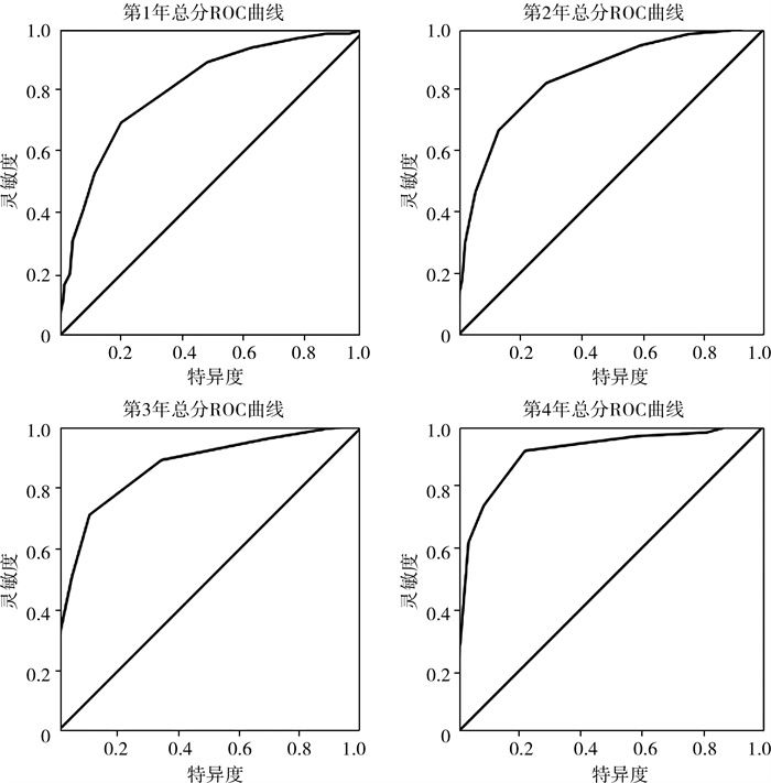
根据本预测模型计算出每个髋关节术后第1、2、3、4年的总分,然后做出第1、2、3、4年的ROC曲线(表 7)。由表 7和图 1可知,本模型术后第1~4年预测最终痊愈的ROC曲线下面积(area under the curve, AUC)为0.808~0.910(图 1)。本模型中,AUC均大于0.8,具有良好的区分度。根据ROC曲线计算出临界总分,由表 7可知,术后第1~4年的临界总分为1.5~2.5分(表 7),当总分小于该值时,痊愈概率显著升高。
| 表 7 DDH患儿闭合复位术后第1、2、3、4年各指标总分的ROC曲线分析 Table 7 ROC curve analysis of total scores of different factors at one to four years post reduction in patients with DDH |
|
|

|
图 1 DDH患儿术后第1~4年各指标总分的ROC曲线图 Fig.1 ROC curve of total scores of different factors at Year 1/2/3/4 post-reduction in DDH children 注 DDH:发育性髋关节发育不良;ROC: 受试者工作特征曲线 |
根据临界总分进行分组,最终分别有80.4%、83.6%、84%、95.5%的患者获得痊愈(表 8)。同时,计算出该模型预测最终痊愈的Kappa系数为0.497~0.645,由此可见,该模型内部具有中等至较强的校准度或一致性。
| 表 8 DDH闭合复位术后第1、2、3、4年时根据临界分数分组与最终结局的关系 Table 8 Relationship of final outcomes and different groups assigned according to cutoff values at Year 1/2/3/4 post-reduction in DDH children |
|
|
病例1(图 2):女性,16.2个月,左侧DDH,术前IHDI分型Ⅲ型,有股骨头骨化核,采用闭合复位石膏固定。术后第1年AI为25.7°,CEA为14°,RI为27%,股骨头无AVN,总分为0+2+(-2)+0+1+1+0=2分(<2.5分),预测痊愈概率为0.897552133。术后第7年X线检查提示髋关节Severin分级为Ⅰ级,患者获得痊愈。

|
图 2 1例女性左侧DDH患儿手术前后X线片 Fig.2 Preopertive and postoperative radiographic films of a girl with left DDH 注 A:术前IHDI分型Ⅲ型,有股骨头骨化核;B:采用闭合复位石膏固定;C:术后第1年总分为2分(<2.5分),预测痊愈概率为0.897552133;D:术后第7年(D)X线检查提示髋关节Severin分级为Ⅰ级 |
病例2(图 3):女,20.2个月,左侧DDH,术前IHDI分型Ⅳ型,有股骨头骨化核,采用闭合复位石膏固定治疗。术后第1年AI 32.5°(1分),CEA 10.7°(1分),RI 31%(1分),有AVN(0分)。术后第1年总分为1+2+(-2)+1+1+1+5=9分(>2.5分),痊愈概率为0.35580903,预计很难获得痊愈,予继续观察。术后第2年AI 33.2°(1分),CEA 7.6°(1分),RI 42%(2分),Ⅱ型AVN(3分),总分为1+2+(-2)+1+1+2+3=7分,痊愈概率为0.227746,痊愈概率很低。术后第6年影像学检查结果为Severin Ⅲ级。

|
图 3 1例女性20.2月龄左侧DDH患儿手术前后X线片 Fig.3 Preopertive and postoperative radiographic films of a 20.2-month-old girl with left DDH 注 A:术前IHDI分型Ⅳ型,有股骨头骨化核;B:采用闭合复位石膏固定;C:术后第1年总分为9分。D:术后第2年总分为7分,预测痊愈概率很低;E:术后第6年X线检查结果为Severin Ⅲ级,患者有RAD。DDH:发育性髋关节脱位 |
目前,国内外对于DDH闭合复位术后结局的预测还存在很多争议。Albinana等[10]回顾性研究了72例采用闭合复位的DDH患者,末次随访时47髋(65%)为Severin Ⅰ、Ⅱ级,25髋(35%)为Severin Ⅲ、Ⅳ级,发现AI可以预测最终结局,复位后2年AI>35°的患者有80%的概率出现Severin Ⅲ、Ⅳ级的结局。Li等[13]回顾性研究了多个中心的89例DDH患者资料,发现AI是预测结局的最优指标,闭合复位术后如果1年AI>28°,2~4年AI>25°,则很可能出现RAD。Fu等[20]回顾性研究了36例(48髋)采用闭合复位治疗的DDH患者资料,术后每年测量AI、CEA、RI,结果显示,如果在3~4岁时RI>38%或4~5岁时RI>33%且髋臼眉弓向上,则很可能出现不满意的结局。
鉴于此,目前国内外对于RAD行骨盆截骨矫形的手术指征和时机存在很大争议。Schwartz[21]回顾性研究了50例采用闭合复位的DDH患者资料,认为DDH闭合复位后2年,如果AI>25°则建议行骨盆截骨术。Shin等[22]则认为,DDH复位3年后,如果AI>32°、CEA<14°则有行骨盆截骨矫形的指征。Tasnavites等[23]认为,二期骨盆截骨矫形应在闭合复位3年以后进行,且AI值要大于正常值的2个标准差。此外,Tönnis[24]、Vrdoljak等[25]、Kim等[26]、Gotoh等[27]、Albinana等[10]、Kitoh等[28]、Terjesen[29]、Fu等[20]、Li[13]也通过研究提出了不同的手术指征和手术时机。他们提出的手术时机从术后2年至术后8~10年不等,手术指征主要依据AI、CEA和RI值,但各研究得出的数值存在较大差距。可见,目前不同研究者对于RAD行骨盆截骨术的手术指征和手术时机存在较大差异。
既往研究中有关DDH闭合复位术后结局的预测,国内外研究主要存在以下缺陷。一是部分研究者得出的结论缺乏数据支持,而是仅凭自己的经验。二是多数研究中评估髋关节影像学表现的指标单一,绝大多数研究只纳入了AI,少部分还包括了CEA、RI。众所周知,髋关节的发育可能受到很多因素的影响,如年龄、脱位严重程度、治疗前AI、股骨头骨化核是否出现、AVN等。仅凭AI或CEA等单一指标很难充分评估髋臼的发育情况。因此,建立一个可以综合多个因素的模型对于预测DDH闭合复位术后结局具有重要意义。
本研究构建了DDH闭合复位后痊愈预测模型。目前,国内外尚无研究者通过构建模型来预测RAD的发生。本研究构建的模型纳入了3个基础指标(复位年龄、复位前的脱位程度IHDI分型和股骨头骨化核是否出现)和4个当前指标(AI、CEA、RI、AVN)。既往研究显示,这些指标都是影响DDH闭合复位术后结局的重要因素[13, 20, 30-33]。利用该模型,我们可以预测DDH闭合复位术后的结局,且具有可操作性。对于DDH闭合复位后4年以内的任意一个时间点,我们获取复位年龄、复位前IHDI分型、有无股骨头骨化核,以及当前AI、CEA、RI、AVN等指标数据,通过该模型便可以得到获得痊愈的概率值。此外,为了进一步指导该预测模型的应用,本模型还确定了是否获得痊愈的临界总分。术后第1年总分<2.5分、术后第2年总分<1.5分、术后第3年总分<2.5分、术后第4年总分<1.5分时,痊愈概率将显著升高。反之则出现RAD的概率显著升高(图 2、图 3)。验证结果显示,当总分小于临界分数时,81.4%~96.1%的患儿可以获得痊愈,可见该模型具有良好的可靠性。
本模型中,AVN是预测模型中权重最大的因素。既往研究也表明,AVN是影响最终影像学结果的重要因素。Malvitz等[34]回顾性研究了119例(152髋)采用闭合复位治疗的DDH患者,平均随访时间达30年,结果显示,有AVN的DDH患者最终影像学和髋关节功能较没有AVN的患者明显变差。Terjesen等[35]也对71例(90髋)DDH患者进行了50年的随访研究,这些患者中有83髋获得稳定复位,末次随访时有27髋(33%)出现骨性关节炎,进一步分析发现,AVN是骨性关节炎的重要风险因素。AVN的出现会严重影响股骨头以及股骨颈的形态,导致股骨头外翻或内翻、扁平、变粗,股骨颈变短,造成股骨头包容欠佳,从而严重影响影像学结果和髋关节功能。
与既往研究采用的预测方法相比,本研究构建的模型具有诸多优点:①本模型综合了多个指标,避免了单一指标的局限性;②该模型可操作性好,基础指标和当前指标都很容易获取;③模型给出了具体的分数和预测痊愈概率值,医师可以更直观地判断患者当前的病情;④本模型提供了多个时间点的预测结果,临床医师可以比较前后多个时间点的预测概率,从而对患者的病情变化进行判断;⑤给出了临界分数,当总分小于等于临界分数时预计可以获得痊愈。因此,本研究制定的预测模型对于DDH闭合复位术后RAD的预测具有重要的临床价值,可以有效地甄别出哪些患者能够获得痊愈,哪些患者可能出现RAD,从而决定是否行骨盆截骨术。当然,本研究也存在不足。由于数据所限,本研究还没有进行外部数据验证。下一步我们将通过中国儿童骨科多中心研究协作组平台,纳入更多病例,对该模型进行修正和外部验证。
综上所述,本研究构建的痊愈预测模型可以有效预测RAD,并指导骨盆截骨手术时机的选择。该模型纳入了复位年龄、复位前IHDI分型、股骨头骨化核是否出现、AI、CEA、RI、AVN等指标。在该模型下,如果术后第1~4年总分小于1.5~2.5分,则很可能获得痊愈;而大于1.5~2.5分,则很可能出现RAD,建议行骨盆截骨矫形。
利益冲突 所有作者声明不存在利益冲突
作者贡献声明 石伟哲、林雪梅负责文献检索,黎艺强、刘行、郭跃明、徐宏文负责论文设计,韦胜、徐晨晨、刘行、郭跃明负责数据收集,黎艺强、刘远忠、徐宏文负责研究结果分析与讨论,黎艺强负责论文撰写;徐宏文负责全文知识性内容的审读与修正
| [1] |
Cooper AP, Doddabasappa SN, Mulpuri K. Evidence-based management of developmental dysplasia of the hip[J]. Orthop Clin North Am, 2014, 45(3): 341-354. DOI:10.1016/j.ocl.2014.03.005 |
| [2] |
Aresti NA, Ramachandran M, Paterson M, et al. Paediatric orthopaedics in clinical practice[M]. London: Springer, 2016. DOI:10.1007/978-1-4471-6769-3
|
| [3] |
Tao ZB, Wang J, Li YM, et al. Prevalence of developmental dysplasia of the hip (DDH) in infants: a systematic review and meta-analysis[J]. BMJ Paediatr Open, 2023, 7(1): e002080. DOI:10.1136/bmjpo-2023-002080 |
| [4] |
Almutairi FF. Incidence and characteristics of developmental dysplasia of the hip in a Saudi population: a comprehensive retrospective analysis[J]. Medicine (Baltimore), 2024, 103(6): e36872. DOI:10.1097/MD.0000000000036872 |
| [5] |
蒋飞, 乔飞, 孙磊娇, 等. 大连地区婴幼儿发育性髋关节发育不良初步筛查及高危因素分析[J]. 临床小儿外科杂志, 2017, 16(2): 159-163, 188. Jiang F, Qiao F, Sun LJ, et al. Preliminary screening of infants with developmental dysplasia of the hip and analysis of risk factors in Dalian[J]. J Clin Ped Sur, 2017, 16(2): 159-163, 188. DOI:10.3969/j.issn.1671-6353.2017.02.013 |
| [6] |
高国朋, 严双琴, 葛群, 等. 马鞍山市市区婴幼儿髋关节发育不良早期筛查[J]. 中国妇幼保健, 2018, 33(1): 124-126. Gao GP, Yan SQ, Ge Q, et al. Early screening of infants with dysplasia of the hip in Maanshan[J]. Matern Child Health Care China, 2018, 33(1): 124-126. DOI:10.7620/zgfybj.j.issn.1001-4411.2018.01.44 |
| [7] |
汤喆滢, 王雯雯, 潘蕾. 天津市10262名婴儿髋关节超声筛查结果及相关因素分析[J]. 中国妇幼保健, 2011, 26(33): 5177-5178. Tang ZY, Wang WW, Pan L. The hip ultrasound screening outome of 10262 infants and analysis of relative factors[J]. Matern Child Health Care China, 2011, 26(33): 5177-5178. |
| [8] |
中华医学会小儿外科分会骨科学组, 中华医学会骨科学分会小儿创伤矫形学组. 发育性髋关节发育不良临床诊疗指南(0~2岁)[J]. 中华骨科杂志, 2017, 37(11): 641-650. Chinese Medical Association Pediatric Surgery Branch Orthopaedic Group, Chinese Medical Association Orthopaedic Branch Pediatric Trauma and Orthopaedic Group. Detection and treatment of pediatric developmental dysplasia of the hip in children up to two year of age: clinical practice guideline[J]. Chin J Orthop, 2017, 37(11): 641-650. DOI:10.3760/cma.j.issn.0253-2352.2017.11.001 |
| [9] |
Li YQ, Lin XM, Liu YH, et al. Effect of age on radiographic outcomes of patients aged 6-24 months with developmental dysplasia of the hip treated by closed reduction[J]. J Pediatr Orthop B, 2020, 29(5): 431-437. DOI:10.1097/BPB.0000000000000672 |
| [10] |
Albinana J, Dolan LA, Spratt KF, et al. Acetabular dysplasia after treatment for developmental dysplasia of the hip.Implications for secondary procedures[J]. J Bone Joint Surg Br, 2004, 86(6): 876-886. DOI:10.1302/0301-620x.86b6.14441 |
| [11] |
Cooperman DR, Wallensten R, Stulberg SD. Acetabular dysplasia in the adult[J]. Clin Orthop Relat Res, 1983, 175: 79-85. |
| [12] |
Narayanan U, Mulpuri K, Sankar WN, et al. Reliability of a new radiographic classification for developmental dysplasia of the hip[J]. J Pediatr Orthop, 2015, 35(5): 478-484. DOI:10.1097/BPO.0000000000000318 |
| [13] |
Li YQ, Guo YM, Li M, et al. Acetabular index is the best predictor of late residual acetabular dysplasia after closed reduction in developmental dysplasia of the hip[J]. Int Orthop, 2018, 42(3): 631-640. DOI:10.1007/s00264-017-3726-5 |
| [14] |
Kalamchi A, MacEwen GD. Avascular necrosis following treatment of congenital dislocation of the hip[J]. J Bone Joint Surg Am, 1980, 62(6): 876-888. DOI:10.2106/00004623-198062060-00002 |
| [15] |
Severin E. Contribution to the knowledge of congenital dislocation of the hip joint[J]. Acta Chir Scand, 1941, 84(Suppl 63): 1-142. |
| [16] |
Gage JR, Winter RB. Avascular necrosis of the capital femoral epiphysis as a complication of closed reduction of congenital dislocation of the hip.A critical review of twenty years' experience at Gillette Children's Hospital[J]. J Bone Joint Surg Am, 1972, 54(2): 373-388. DOI:10.2106/00004623-197254020-00015 |
| [17] |
石永言, 刘天婧, 赵群, 等. 中国人髋关节髋臼指数和Sharp角正常值的测量[J]. 中华骨科杂志, 2010, 30(8): 748-753. Shi YY, Liu TJ, Zhao Q, et al. The measurements of normal acetabular index and Sharp acetabular angle in Chinese hips[J]. Chin J Orthop, 2010, 30(8): 748-753. DOI:10.3760/cma.j.issn.0253-2352.2010.08.004 |
| [18] |
刘远忠, 郭跃明, 沈先涛, 等. 牵引在闭合复位治疗儿童发育性髋关节脱位中的作用的多中心回顾性研究[J]. 中华小儿外科杂志, 2017, 38(7): 500-505. Liu YZ, Guo YM, Shen XT, et al. Effect of traction on developmental dysplasia of the hip treated via closed reduction: a multi-center retrospective study[J]. Chin J Pediatr Surg, 2017, 38(7): 500-505. DOI:10.3760/cma.j.issn.0253-3006.2017.07.004 |
| [19] |
D'Agostino RB Sr, Grundy S, Sullivan LM, et al. Validation of the framingham coronary heart disease prediction scores: results of a multiple ethnic groups investigation[J]. JAMA, 2001, 286(2): 180-187. DOI:10.1001/jama.286.2.180 |
| [20] |
Fu Z, Yang JP, Zeng P, et al. Surgical implications for residual subluxation after closed reduction for developmental dislocation of the hip: a long-term follow-up[J]. Orthop Surg, 2014, 6(3): 210-216. DOI:10.1111/os.12113 |
| [21] |
Schwartz DR. Acetabular development after reduction of congenital dislocation of the hip: a follow-up study of fifty hips[J]. J Bone Joint Surg Am, 1965, 47: 705-714. DOI:10.2106/00004623-196547040-00005 |
| [22] |
Shin CH, Yoo WJ, Park MS, et al. Acetabular remodeling and role of osteotomy after closed reduction of developmental dysplasia of the hip[J]. J Bone Joint Surg Am, 2016, 98(11): 952-957. DOI:10.2106/JBJS.15.00992 |
| [23] |
Tasnavites A, Murray DW, Benson MK. Improvement in acetabular index after reduction of hips with developmental dysplasia[J]. J Bone Joint Surg Br, 1993, 75(5): 755-759. DOI:10.1302/0301-620X.75B5.8376433 |
| [24] |
Tönnis D. Congenital dysplasia and dislocation of the hip in children and adults[M]. Berlin: Springer-Verlag, 1987. DOI:10.1007/978-3-642-71038-4
|
| [25] |
Vrdoljak J, Gogolja D. Development of acetabulum after closed reduction in developmental hip dysplasia[J]. Coll Antropol, 1999, 23(2): 745-749. |
| [26] |
Kim HT, Kim JI, Yoo CI. Acetabular development after closed reduction of developmental dislocation of the hip[J]. J Pediatr Orthop, 2000, 20(6): 701-708. DOI:10.1097/00004694-200011000-00002 |
| [27] |
Gotoh E, Tsuji M, Matsuno T, et al. Acetabular development after reduction in developmental dislocation of the hip[J]. Clin Orthop Relat Res, 2000, 378: 174-182. DOI:10.1097/00003086-200009000-00027 |
| [28] |
Kitoh H, Kitakoji T, Katoh M, et al. Prediction of acetabular development after closed reduction by overhead traction in developmental dysplasia of the hip[J]. J Orthop Sci, 2006, 11(5): 473-477. DOI:10.1007/s00776-006-1049-2 |
| [29] |
Terjesen T. Residual hip dysplasia as a risk factor for osteoarthritis in 45 years follow-up of late-detected hip dislocation[J]. J Child Orthop, 2011, 5(6): 425-431. DOI:10.1007/s11832-011-0370-2 |
| [30] |
Li YQ, Liu H, Guo YM, et al. Variables influencing the pelvic radiological evaluation in children with developmental dysplasia of the hip managed by closed reduction: a multicentre investigation[J]. Int Orthop, 2020, 44(3): 511-518. DOI:10.1007/s00264-020-04479-z |
| [31] |
Terjesen T. Long-term outcome of closed reduction in late-detected hip dislocation: 60 patients aged six to 36 months at diagnosis followed to a mean age of 58 years[J]. J Child Orthop, 2018, 12(4): 369-374. DOI:10.1302/1863-2548.12.180024 |
| [32] |
Eamsobhana P, Saisamorn K, Sisuchinthara T, et al. The factor causing poor results in late Developmental Dysplasia of the Hip (DDH)[J]. J Med Assoc Thai, 2015, 98(Suppl 8): S32-S37. |
| [33] |
Cummings JL, Oladeji AK, Rosenfeld S, et al. Severity of hip dysplasia as the major factor affecting outcome of closed reduction in children with hip dysplasia[J]. J Pediatr Orthop B, 2024, 33(4): 322-327. DOI:10.1097/BPB.0000000000001122 |
| [34] |
Malvitz TA, Weinstein SL. Closed reduction for congenital dysplasia of the hip.Functional and radiographic results after an average of thirty years[J]. J Bone Joint Surg Am, 1994, 76(12): 1777-1792. DOI:10.2106/00004623-199412000-00004 |
| [35] |
Terjesen T, Horn J, Gunderson RB. Fifty-year follow-up of late-detected hip dislocation: clinical and radiographic outcomes for seventy-one patients treated with traction to obtain gradual closed reduction[J]. J Bone Joint Surg Am, 2014, 96(4): e28. DOI:10.2106/JBJS.M.00397 |
 2024, Vol. 23


