2. 中南大学湘雅医学院附属儿童医院泌尿外科, 长沙 410007;
3. 中南大学湘雅医学院附属儿童医院放射科, 长沙 410007
2. Department of Urology, Children's Hospital, Xiangya School of Medicine, Central South University, Changsha 410007, China;
3. Department of Radiology, Children's Hospital, Xiangya School of Medicine, Central South University, Changsha 410007, China
尿石症是小儿泌尿外科常见疾病,病因主要包括遗传代谢异常、生理畸形及尿路感染等,临床症状表现为肾绞痛、血尿、反复尿路感染、尿痛、排尿困难等[1]。目前,小儿泌尿系结石的手术治疗方法主要包括体外冲击波碎石术、经皮肾镜碎石取石术、输尿管镜碎石取石术[2]。刘李等[3]报道,小儿泌尿系结石成分主要包括草酸钙结石(最多见)、尿酸结石、鸟粪石(感染性结石)、胱氨酸结石、混合性结石等,且较成人更易复发[4]。鉴于CT是诊断小儿泌尿系结石灵敏度最高的影像学方法,Rodríguez-Plata等[5]和舒露[6]分别研究报道,可以通过CT豪恩斯菲尔德单位(hounsfield unit, HU)预测结石成分,为泌尿系结石患者的诊疗提供依据。本研究旨在探讨小儿泌尿系结石CT HU值在结石成分判断中的应用价值。
资料与方法 一、研究对象本研究为回顾性研究,以2009年4月至2022年4月在湖南省儿童医院诊断为泌尿系结石的患儿为研究对象。病例纳入标准:①首次在本院行碎石取石手术治疗;②术前在本院行CT影像学检查;③术中或术后获取结石标本,且术后结石成分单一。排除标准:①结石长径(结石在横断面上最大横截面积的最长尺寸)≤3 mm;②某种单一结石成分类型病例总数<10例(如黄嘌呤结石5例)。根据上述标准,最终纳入本研究的泌尿系结石患儿共422例。本研究经过湖南省儿童医院伦理委员会审核批准(HCHLL-2022-137),患儿父母均知情同意。
二、数据的收集CT HU值测量:患儿治疗前均接受了泌尿系CT扫描影像学检查[检查设备:飞利浦64排螺旋CT(型号:PHILIPS CT Brilliance),使用3 mm薄层厚度。进入医疗影像系统(picture archiving and communication system, PACA),在CT平扫骨窗界面,结石长径大于3 mm认为有临床诊断意义。由CT扫描图像中结石长径层面的感兴趣区(region of interest, ROI)测量结石的CT HU值。在结石长径中心测量一次,标记为结石核心HU值;在结石长径外围分别测量两次,测量的HU值平均数标记为结石外围HU值;
采用智能结石分析仪(型号:SUN-3G,济南鼎舜医疗器械有限公司)进行结石成分分析。红外光谱结石成分分析流程:①首先将结石标本用蒸馏水洗干净,放入70℃ ~100℃烤箱内烘干待用;②将结石样本研磨成粉末;③将纯溴化钾进行干燥处理,取200 mg干燥溴化钾与1 mg结石粉末样本混合,在玛瑙研钵中碾压成粉末充分混合;④将结石标本置于压片机中制备成半透明状检测片;⑤将检测片植入光谱槽中扫描,自动分析出结石成分。根据结石成分分析结果,将所有患儿分为4组:草酸钙结石组、尿酸结石组、鸟粪石组、胱氨酸结石组。草酸钙结石包括一水草酸钙和(或)二水草酸钙;尿酸结石包括无水尿酸、尿酸氢铵、一水尿酸;鸟粪石即感染性结石,包括磷酸铵镁和(或)碳酸磷灰石。
三、统计学处理采用SPSS 25.0进行统计学分析。对服从正态分布的计量资料采用x±s表示;对多个符合正态分布的样本均数两两比较使用Dunnett-t检验进行方差分析,P<0.05为差异有统计学意义。计数资料以频数、构成比表示,不满足正态分布的变量资料使用[M(Q1,Q3)]表示;多组间两两比较采用多样本的秩和检验(Kruskal-Wallis Dunn test),基于两两比较的秩次,显著性水平设定为P<0.008为差异有统计学意义。采用受试者工作特征曲线(receiver operating curve, ROC)分别计算各组参数截断值、ROC曲线下面积(area under curve, AUC)等。
结果本研究纳入的422例患儿中,年龄范围为4~204个月,平均年龄为67.4个月;男性284例(67.3%),女性138例(32.7%);草酸钙结石组273例,尿酸结石组89例,鸟粪石组38例,胱氨酸结石组22例,占比分别为65%、21%、9%、5%。
4组患儿结石外围HU值和结石平均HU值:尿酸结石组较草酸钙结石组、鸟粪石组、胱氨酸结石组均小(P<0.05)。4组结石核心HU值:尿酸结石组较草酸钙结石组和鸟粪石组小(P<0.05)。4组HUD:草酸钙结石组较尿酸结石组、鸟粪石组、胱氨酸结石组大(P<0.05);尿酸结石组较鸟粪石组小(P<0.05)。4组中位尿pH值:胱氨酸结石组较草酸钙结石组和尿酸结石组大(P<0.001)。见表 1。
| 表 1 4组不同成分泌尿系结石患儿CT HU值、尿pH值比较 Table 1 Comparing four groups of different calculus composition with CT HU value and urinary pH |
|
|
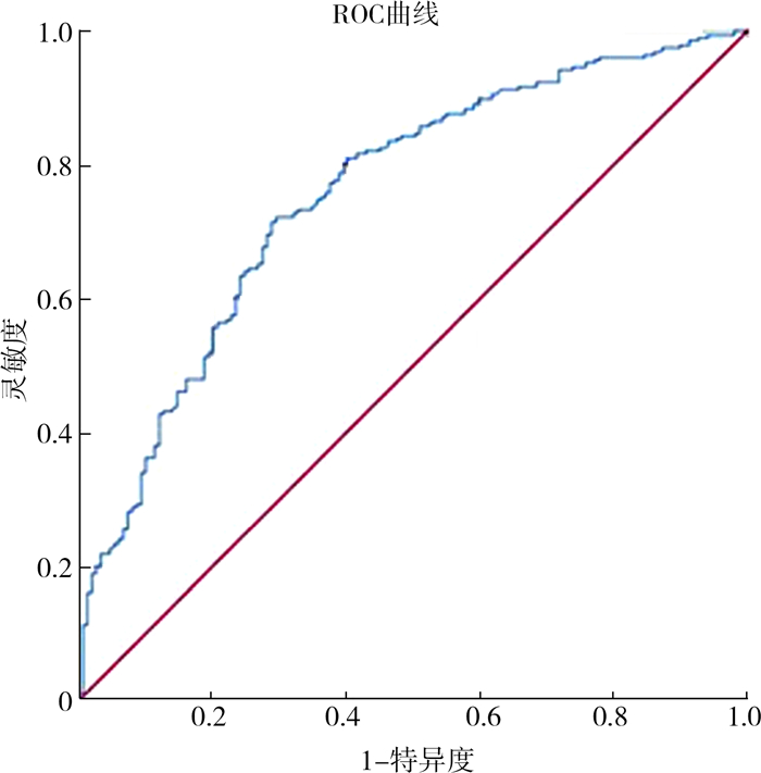
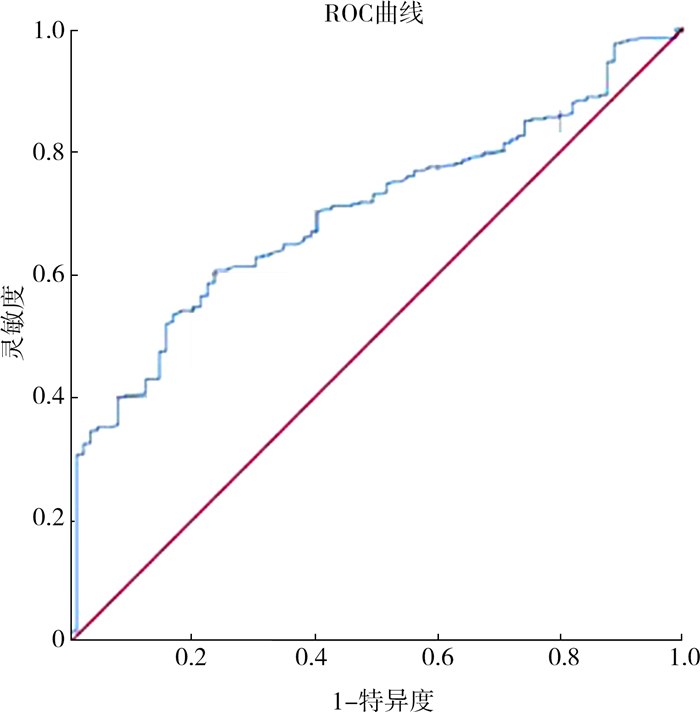
草酸钙结石组与非草酸钙结石组HUD的AUC为0.755,灵敏度为0.722,特异度为0.705,最佳截断值为48;非尿酸结石组与尿酸结石组的核心HU值AUC为0.700,灵敏度为0.607,特异度为0.764,最佳截断值为566;胱氨酸结石组与非胱氨酸结石组中位尿pH值AUC为0.704,灵敏度为0.682,特异度为0.657,最佳截断值为6.25。详见图 1至图 3、表 2至表 4。

|
图 1 草酸钙结石与非草酸钙结石HUD参数的ROC曲线下面积 Fig.1 Area under the ROC curve for HUD parameter of calcium oxalate and non-calcium oxalate calculi |

|
图 2 非尿酸结石与尿酸结石的结石核心HU值参数ROC曲线下面积 Fig.2 Area under the ROC curve of HU value parameters in core of non-uric acid and uric acid calculi |

|
图 3 胱氨酸结石与非胱氨酸结石尿pH值参数ROC曲线下面积 Fig.3 Area under the ROC curve of urinary pH parameters between cystine and non-cystine calculi |
| 表 2 HUD鉴别草酸钙结石成分诊断价值分析 Table 2 Diagnostic value of HUD in the identification of calcium oxalate calculus |
|
|
| 表 3 结石核心HU值鉴别非尿酸结石成分诊断价值分析 Table 3 Core HU diagnostic values for identifying uric acid calculus compositions |
|
|
| 表 4 尿pH值鉴别胱氨酸结石成分诊断价值分析 Table 4 Diagnostic value of urinary pH for identifying cystine calculi ingredients |
|
|
小儿泌尿系结石的成分与其临床治疗、预后有关,不同的结石成分、大小、位置均会直接关系到患儿的治疗效果。小儿泌尿系结石成分包括草酸钙结石、尿酸结石、鸟粪石、胱氨酸结石、混合性结石等。2022年,刘李等[3]报道了592例儿童泌尿系结石成分分析结果,单一结石成分以草酸钙结石、尿酸氢铵为主,分别占63.7%、14.3%。本研究中单一结石成分的草酸钙结石、尿酸结石、鸟粪石、胱氨酸结石分别占65%、21%、9%、5%,与文献报道结果相符。邢滇霞[7]于2017年报道,胱氨酸结石在体外冲击波碎石取石中难度最大,草酸钙结石的碎石难度相对较大,鸟粪石的碎石难度最低。鉴于CT影像学检查是诊断泌尿系结石最敏感的方法,不仅快速、精准、全面,还可通过CT HU值的测量预测患儿结石成分。因此在泌尿系结石患儿的治疗过程中,术前通过CT扫描识别结石成分类型,选择合适的治疗方法,对改善患儿的治疗效果有重要意义。
本研究发现,草酸钙结石组HUD高于尿酸结石组、鸟粪石组、胱氨酸结石组;尿酸结石组的结石外围HU值、结石核心HU值、结石平均HU值、HUD均低于其他三组。当HUD大于等于48时,提示草酸钙结石;当结石核心HU值小于566时,提示尿酸结石;当尿pH值大于等于6.25时,提示胱氨酸结石;排除其他结石成分后,其余结石成分类型提示为鸟粪石。这与Altan等[8]研究报道草酸钙结石核心CT HU值、外围CT HU值、平均CT HU值和HUD的样本均数均高于胱氨酸结石和鸟粪石的结果有一定差别。在区分胱氨酸结石和鸟粪石时,CT HU值、HUD比较均无明显差异,需要引入尿pH值等参数。依据胱氨酸结石组与非胱氨酸结石组ROC曲线分析结果,当尿pH值截断值设定在(6.25)时,灵敏度、特异度、准确度较高,对于区分胱氨酸结石和非胱氨酸结石有一定的参考意义。这一发现与Altan等[8]报道的鸟粪石组的尿pH值中位数高于胱氨酸组有差别,胱氨酸结石患儿治疗过程中需要碱化尿液。临床工作中还需参考患儿的血常规、C反应蛋白、降钙素原、尿常规白细胞数、尿培养等感染性指标,进一步区分胱氨酸结石和鸟粪石。
对于鸟粪石(感染性结石)患儿,因结石大部分发生在肾脏,生长速度快,常迅速填满肾盂和肾大盏,从而导致患儿肾功能丧失,易并发严重感染,结石复发率和致死率均较高[9]。Schnabel等[10]认为,抗感染治疗对于感染性结石的防治具有重要的作用,未经治疗的感染性结石是泌尿外科微创手术的禁忌。术前通过CT HU值等参数判断患儿结石成分,如为感染性结石,则采取积极干预措施,在围手术期使用抗生素进行抗感染治疗,术中相对缩短手术时间,保持较低的肾盂内灌注压,从而减少围手术期全身炎症反应、脓毒血症、肾功能衰竭、感染性休克等感染相关并发症的发生[11]。胱氨酸结石主要是由于胱氨酸尿石症所致,具有遗传特异性,也被称为遗传性结石。赵夭望等[12]研究报道,胱氨酸结石患儿术后复发率极高,且胱氨酸结石质韧,体外冲击波碎石通常不易粉碎。如术前相关指标提示为胱氨酸结石,则可选择除体外冲击波碎石以外的治疗方法,进而提高临床治疗效果。
CT HU值与泌尿系结石成分的相关性研究多以成人为研究对象,这些研究中使用了结石平均CT HU值、最大CT HU值、最小CT HU值、结石异质性指数、平均结石密度、双能量指数等参数预测结石成分[13]。因为结石的大小与CT HU值呈正相关,为避免结石大小的影响,本研究中同样使用了HUD参数[14]。而通过CT HU值参数,很难判断鸟粪石和胱氨酸结石成分,因此引入了尿pH值这一参数[8]。在相关研究中,结石异质性指数、平均结石密度、双能量指数等参数对于预测结石成分很有效,但与结石核心CT HU值、外围CT HU值、HUD等相比,并没有突出的优势[15]。CT检查诊断泌尿系结石的灵敏度、特异度都很高,大部分患儿不需要镇静,可快速完成检查,明确病情,且在评估泌尿系结石患儿的急性输尿管结石症状、结石负担、已知泌尿系生殖道畸形和可疑结石症状等方面有明显的优势[16]。但根据Lam等[17]研究报道,CT扫描仍存在一定辐射,尽管辐射量较低,但如果反复行CT检查,会导致一定的累积辐射暴露,特别是胱氨酸结石患儿,术后易复发。因此,小儿泌尿系结石患儿术后随访中,可优先考虑泌尿系彩超检查。
本研究的局限性在于为单中心研究,样本量有限。同时,结石成分类型按照单一结石成分分组,但并不包括一些罕见的单一结石成分类型(如黄嘌呤结石等)。另外,临床中部分患儿结石成分类型为混合性结石成分,对于此类结石成分的术前判断有待进一步研究。
综上所述,通过CT HU值测量,能为判定草酸钙结石和尿酸结石成分提供参考,结石核心HU值小于566时,提示尿酸结石;HUD大于等于48时,提示草酸钙结石;胱氨酸结石成分和鸟粪石结石成分的进一步判断,需要参考尿pH值等参数。
利益冲突 所有作者声明不存在利益冲突
作者贡献声明 覃锋、何天衢、赵夭望负责研究的设计、实施和起草文章;李创业、易婷、孙小文、刘畅、李艳芳、邹志敏进行病例数据收集和分析;赵夭望、王智负责研究设计与酝酿,并对文章知识性内容进行审阅
| [1] |
Magni G, Unwin RJ, Moochhala SH. Renal tubular acidosis (RTA) and kidney stones: diagnosis and management[J]. Arch Esp Urol, 2021, 74(1): 123-128. |
| [2] |
Kovacevic L. Diagnosis and management of nephrolithiasis in children[J]. Pediatr Clin North Am, 2022, 69(6): 1149-1164. DOI:10.1016/j.pcl.2022.07.008 |
| [3] |
刘李, 彭柳成, 李创业, 等. 单中心592例儿童泌尿系结石成分分析[J]. 中华泌尿外科杂志, 2022, 43(9): 701-706. Liu L, Peng LC, Li CY, et al. Analysis of urinary calculi composition of 592 children at a single center[J]. Chin J Urol, 2022, 43(9): 701-706. DOI:10.3760/cma.j.cn112330-20201125-00788 |
| [4] |
Ye ZQ, Zeng GH, Yang H, et al. The status and characteristics of urinary stone composition in China[J]. BJU Int, 2020, 125(6): 801-809. DOI:10.1111/bju.14765 |
| [5] |
Rodríguez-Plata IT, Medina-Escobedo M, Basulto-Martínez M, et al. Implementation of a technique based on Hounsfield units and Hounsfield density to determine kidney stone composition[J]. Tomography, 2021, 7(4): 606-613. DOI:10.3390/tomography7040051 |
| [6] |
舒露. CT值在感染性结石中的初步研究及预警[D]. 遵义: 遵义医科大学, 2020. Shu L. Preliminary researches and early warnings of CT value in infectious calculi[D]. Zunyi: Zunyi Medical University, 2020. |
| [7] |
邢滇霞. 能谱CT在体分析泌尿系结石成分与碎石难易程度的关系[D]. 石河子: 石河子大学, 2017. Xing DX. Energy spectrum CT in vivo analysis of urinary calculi composition and its relationship with grinding difficulties[D]. Shihezi: Shihezi University, 2017. |
| [8] |
Altan M, Çitamak B, Bozaci AC, et al. Predicting the stone composition of children preoperatively by Hounsfield unit detection on non-contrast computed tomography[J]. J Pediatr Urol, 2017, 13(5): 505.e1-505.e6. DOI:10.1016/j.jpurol.2017.03.013 |
| [9] |
Senocak C, Ozcan C, Sahin T, et al. Risk factors of infectious complications after flexible uretero-renoscopy with laser lithotripsy[J]. Urol J, 2018, 15(4): 158-163. DOI:10.22037/uj.v0i0.3967 |
| [10] |
Schnabel MJ, Wagenlehner FME, Schneidewind L. Perioperative antibiotic prophylaxis for stone therapy[J]. Curr Opin Urol, 2019, 29(2): 89-95. DOI:10.1097/MOU.0000000000000576 |
| [11] |
Tekgül S, Stein R, Bogaert G, et al. European Association of Urology and European Society for Paediatric Urology guidelines on paediatric urinary stone disease[J]. Eur Urol Focus, 2022, 8(3): 833-839. DOI:10.1016/j.euf.2021.05.006 |
| [12] |
赵夭望, 李创业. 儿童遗传性肾结石的治疗进展[J]. 临床小儿外科杂志, 2020, 19(8): 666-671. Zhao YW, Li CY. Recent advances in the treatment of hereditary nephrolithiasis in children[J]. J Clin Ped Sur, 2020, 19(8): 666-671. DOI:10.3969/j.issn.1671-6353.2020.08.002 |
| [13] |
杨斌, 汪道琦, 周元, 等. CT和AI技术预测泌尿系结石成分的研究进展[J]. 临床泌尿外科杂志, 2023, 38(2): 139-145. Yang B, Wang DQ, Zhou Y, et al. Research advances of CT and AI technology in predicting the composition of urinary calculi[J]. J Clin Urol, 2023, 38(2): 139-145. DOI:10.13201/j.issn.1001-1420.2023.02.013 |
| [14] |
Kachroo N, Jain R, Maskal S, et al. Can CT-based stone impaction markers augment the predictive ability of spontaneous stone passage?[J]. J Endourol, 2021, 35(4): 429-435. DOI:10.1089/end.2020.0645 |
| [15] |
Yu J, Zhou QC, Lin F, et al. Performance of dual-source CT in calculi component analysis: a systematic review and meta-analysis of 2151 calculi[J]. Can Assoc Radiol J, 2021, 72(4): 742-749. DOI:10.1177/0846537120951992 |
| [16] |
Zumstein V, Betschart P, Hechelhammer L, et al. CT-calculometry (CT-CM): advanced NCCT post-processing to investigate urinary calculi[J]. World J Urol, 2018, 36(1): 117-123. DOI:10.1007/s00345-017-2092-7 |
| [17] |
Lam JP, Alexander LF, William HE, et al. In vivo comparison of radiation exposure in third-generation vs second-generation dual-source dual-energy CT for imaging urinary calculi[J]. J Endourol, 2021, 35(11): 1581-1585. DOI:10.1089/end.2021.0103 |
 2024, Vol. 23


