2. 重庆医科大学附属第一医院骨科, 重庆 400015;
3. 重庆医科大学附属第一医院康复科, 重庆 400015;
4. 重庆医科大学附属儿童医院骨科, 重庆 400015
2. Department of Orthopedics, First Affiliated Hospital, Chongqing Medical University, Chongqing 400015, China;
3. Department of Rehabilitation, First Affiliated Hospital, Chongqing Medical University, Chongqing 400015, China;
4. Affiliated Children's Hospital, Chongqing Medical University, Chongqing 400015, China
足弓是由内侧纵弓、外侧纵弓及横弓组成的三维缓冲系统,在运动中起着重要作用[1]。足部负重时,重力通过足弓向前后、内外侧传递,进而提高负重时的稳定性。在跑步运动中,足弓承担着减轻地面反作用力对关节及软组织冲击力的作用。然而,足弓形态存在个体差异,不同类型足弓存在生物力学的不同,影响跑步过程中地面冲击力的衰减程度及足底压力的分布, 足弓形态变化对于体育运动有着重要影响[2-5]。
多项研究表明,足弓发育异常会改变下肢负荷,导致下肢关节活动度下降。Williams等[6]研究发现,高弓足所致关节活动度降低、下肢负荷增加,可能降低运动能力。Zhao等[7]分别于30°/s和120°/s的角速度下测量不同足型踝关节跖屈、背屈、外翻、内翻的峰值扭矩发现,扁平足患者踝关节扭矩较高,而高弓足患者踝关节扭矩较低,以上异常均可能对运动能力造成影响。因此,足弓的正常发育是运动能力的重要保障。儿童足部处于快速发育时期,关注足弓发育对于儿童运动能力的正常发展具有重要意义。
然而,目前关于儿童足弓形态与运动能力的关系仍然存在争议。Tudor等[8]通过比较扁平足儿童与正常足儿童发现,扁平足儿童运动能力与正常足儿童相当。但Chen等[9]研究发现,扁平足儿童出现运动发育迟缓的概率较正常足儿童高出50%左右,严重影响儿童运动能力的发展。出现上述差异的原因可能是各项实验采取了不同的参数来评价足弓形态,如足弓高度、足印分析法中的足弓指数等二维参数[10-12]。足弓是一个三维动态结构,通过其三维形态变化,储存和释放足部负重时的弹性势能[1]。如使用二维参数则无法准确反映足弓动态变化情况[13-14]。因此,Zhao等[15]提出了一项检测足弓三维指标即足弓体积的方法,该指标不仅可以精准反映儿童足部形态,还可以对足弓动态变化进行描述。相比现有的足弓检测方法(舟骨高度、足弓面积),具有较好的灵敏度和有效性。本研究针对7~15岁特定人群进行足弓形态评价,探讨足弓形态与运动能力的关系。研究中对于不同年龄段儿童的运动能力,根据《国家学生体质健康标准》(2014年修订)要求,以50米短距离奔跑(以下简称短跑)成绩作为标准[16];对于儿童足弓形态,选择传统二维指标“足弓指数”与三维指标“足弓体积”进行统计,分析足弓形态与运动能力的相关性。
资料与方法 一、研究对象本研究为回顾性研究。以重庆市某中小学7~15岁学生为研究对象。纳入标准:①未经过系统体育训练。②身体健康,无遗传病史,无明显足部外伤。排除标准:①存在足部疾病或畸形。②急性病患者或1个月内有过高烧、腹泻等,体力尚未恢复者。③曾接受足部手术。受试者测试前均需告知家长,并填写知情同意书。本研究获得重庆医科大学附属第一医院伦理委员会审核批准(2020—102)并全程监督研究。
本研究最终共纳入有效研究对象640人,研究之前共招募7~15岁青少年650人(男女各325人),其中4人近期有高烧、腹泻,2人存在发育迟缓,4人未完成测试,被排除在外。
受试者身高和体重测量通过标准化设备及环境进行。使用统一且经过校准的机械式身高坐高计,两人一组(一人测量,一人记录)进行身高测量。读数时,检测人员双眼与水平压板平面等高,以cm为单位,读数精确到小数点后两位。体重测量使用经过校准的电子或机械体重秤,受试者需穿着内衣或轻便衣物,并确保口袋中无物品。在确保秤归零后受试者站在秤的中央,双脚分开平均分配体重。所有测量结果均保留至小数点后两位。
受试者640人身高、体重均在《中国儿童生长标准2005》所示正常范围内[17]。受试者人口学统计数据见表 1。
| 表 1 各年龄组受试者人数及身高、体重及BMI比较(n=640) Table 1 Participant demographics, including number of subjects, height, weight and BMI by age group(n=640) |
|
|
在50米短跑测试之前,受试者在同一体育教练指导下进行热身运动,并伸展腿部和足部肌肉。所有测试均在专业体育教练指导下进行。50米短跑测试方法与要求:7人一组,经体育教练指导后统一采用站立式起跑快速跑至终点。计时员见起跑信号即开始计时,受试者躯干到达终点线垂直面的瞬间停表,计时结束。50米短跑测量结果保留至小数点后两位[18]。
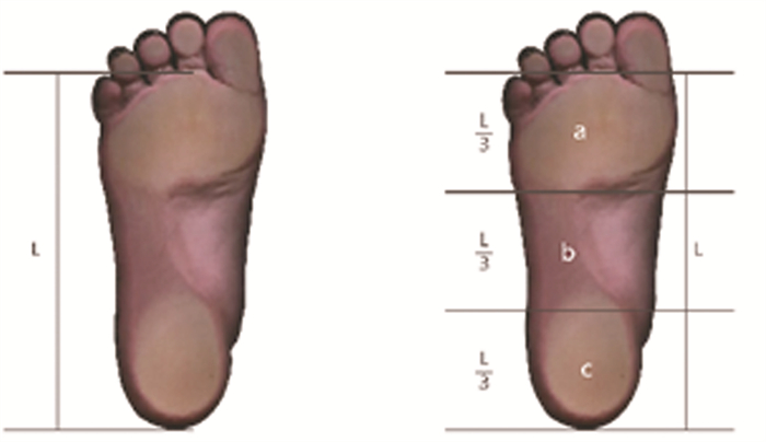
三、足弓指数以及足弓体积的测量使用善启(重庆)智慧医疗科技有限公司制造的“Foot Secret”扫描三维足部轮廓[19]。分别在非负重与负重状态下对受试者进行两次足部扫描。非负重状态下,受试者坐在可调高度的椅子上,保持髋关节和膝关节弯曲90°,踝关节处于中立位置。将双足第二脚趾对准设备激光轴进行定位。扫描后获得足部参数即足弓指数、足弓体积。足弓指数定义为中足面积除以足部面积(不包括脚趾)(图 1)。足弓体积定义为足弓投影到地面的区域与足弓之间的立体空间[20-21](图 2)。

|
图 1 足弓指数示意图 Fig.1 Foot arch index diagram 注 沿L(除脚趾之外的足部长度)将足部分为三等份,中足区域(b)面积与前中后足(a+b+c)区域面积的比值即为足弓指数 |

|
图 2 不同负重条件下足弓体积示意图 Fig.2 Schematic diagram of arch volume under different weight-bearing conditions 注 A:非负重状态下;B:负重状态下 |
采用SPSS 22.0进行数据分析。计数资料用频数、构成比表示。在进行分析之前,使用Shapiro-Wilk对计量资料进行正态性检验。对于服从正态分布的计量资料采用x±s表示,组间比较采用两独立样本t检验;对不服从正态分布的计量资料采用M(Q1, Q3)表示,组间比较采用秩和检验。两组均服从正态分布的计量资料采用Pearson相关性分析;任一组不服从正态分布的计量资料采用Spearman秩相关分析。对于服从正态且方差齐性的不同年龄组的比较采用单因素方差分析,两两比较采用Student-Newman-Keuls Test;对不服从正态分布的不同年龄组的比较采用Kruskal-Wallis检验,两两比较采用Bonfferoni法。P < 0.05为差异有统计学意义。
结果 一、受试者50米短跑成绩不同年龄及性别受试者50米短跑成绩见表 2。随着年龄增长,男女50米短跑成绩均呈上升趋势。其中,男子组由7~9岁时的9.97秒提升至7.99秒,减少1.98秒。女子组由7~9岁时10.29秒提升至9.17秒,减少1.12秒;随着年龄增长,男子组50米短跑成绩提升幅度优于女子组。各年龄阶段,男子组50米短跑成绩均优于女子组,各组差异均有统计学意义(P<0.05)。
| 表 2 不同年龄及性别受试者的50米短跑成绩 Table 2 50-meter sprint results for participants of different age/gender groups |
|
|
在7~9岁中,男子组左足负重足弓指数、左足非负重足弓指数、右足负重足弓指数、右足非负重足弓指数,以及女子组左足负重足弓指数、左足非负重足弓指数、右足负重足弓指数、右足非负重足弓指数与50米短跑成绩呈极弱相关性(r=0.164、0.165、0.189、0.067、0.048、0.049、0.185、0.079),相关系数均无统计学意义(P>0.05)。除女子组左足负重足弓体积、非负重足弓体积表现出极弱相关性(r=-0.173、-0.191),且相关系数无统计学意义(P>0.05)外,男子组左足负重足弓体积、左足非负重足弓体积、右足负重足弓体积、右足非负重足弓体积,以及女子组右足负重足弓体积、右足非负重足弓体积与50米短跑成绩均表现出弱相关性(r=-0.301、-0.294、-0.295、-0.207、-0.279、-0.278),相关系数均有统计学意义(P<0.05),见表 3、表 4。
| 表 3 7~9岁男子组50米短跑成绩与足部参数的相关性分析 Table 3 Correlation analysis between 50-meter sprint results and foot index for boys aged 7-9 |
|
|
| 表 4 7~9岁女子组50米短跑成绩与不同负重状态下足部参数的相关性分析 Table 4 Correlation analysis between 50-meter sprint results and foot index for girls aged 7-9 |
|
|
在10~12岁组中,除女子组右足负重足弓指数、右足非负重足弓指数与50米成绩呈弱相关性(r=0.250、0.243)且相关系数有统计学意义(P<0.05)外,男子组左足负重足弓指数、左足非负重足弓指数、右足负重足弓指数、右足非负重足弓指数,以及女子组左足负重足弓指数、左足非负重足弓指数与50米短跑成绩均呈极弱相关性(r=0.098、0.089、0.105、0.084、0.142、0.142),且相关系数均无统计学意义(P>0.05);除女子组左足负重足弓体积、左足非负重足弓体积与50米短跑成绩呈极弱相关性(r=-0.096、0.077)且相关系数无统计学意义(P>0.05)外,男子组左足负重足弓体积、左足非负重足弓体积、右足负重足弓体积、右足非负重足弓体积,以及女子组右足负重足弓体积、右足非负重足弓体积与50米短跑成绩呈弱相关性(r=-0.273、-0.224、-0.236、-0.273、-0.224、-0.211),且相关系数均有统计学意义(P<0.05)。
四、13~15岁组50米短跑成绩与足部参数在13~15岁组中,男子组左足负重足弓指数、左足非负重足弓指数、右足负重足弓指数、右足非负重足弓指数,以及女子组左足负重足弓指数、左足非负重足弓指数、右足负重足弓指数、右足非负重足弓指数与50米短跑成绩均呈极弱相关性(r= 0.038、-0.025、0.036、-0.096、0.054、0.058、0.031、0.083),且相关系数无统计学意义(P>0.05);除女子组左足非负重足弓体积、右足非负重足弓体积与50米短跑成绩呈弱相关性(r=-0.233、-0.225)且相关系数具有统计学意义(P<0.05)外,男子组左足负重足弓体积、左足非负重足弓体积、右足负重足弓体积、右足非负重足弓体积,以及女子组左足负重足弓体积、右足负重足弓体积与50米短跑成绩呈极弱相关性(r=-0.117、-0.034、-0.131、-0.105、-0.126、-0.128),且相关系数均无统计学意义(P>0.05),见表 7、表 8。
| 表 5 10~12岁男子组50米短跑成绩与不同负重状态下足部参数的相关性分析 Table 5 Correlation analysis between 50-meter sprint results and foot index for boys aged 10-12 |
|
|
| 表 6 10~12岁女子组50米短跑成绩与不同负重状态下足部参数相关性分析 Table 6 Correlation analysis between 50-meter sprint results and foot index for girls aged 10-12 |
|
|
| 表 7 13~15岁男子组50米短跑成绩与不同负重状态下足部参数的相关性分析 Table 7 Correlation analysis between 50-meter sprint results and foot index for boys aged 13-15 |
|
|
| 表 8 13~15岁女子组50米短跑成绩与不同负重状态下足部参数的相关性分析 Table 8 Correlation analysis between 50-meter sprint results and foot index for girls aged 13-15 |
|
|
人体足部是一个高度灵活的多关节结构,通过足部众多关节和软组织之间的协同作用,有效传递下肢与地面之间的力量。足弓和足底筋膜形成了一个封闭的铰链结构,能够完整地吸收和缓冲张力及应力[22]。在站立相前中期,足弓被拉长并压缩,并将冲击力转化为弹性势能吸收。而在站立相末期,地面反作用力使足趾背屈,足底筋膜被拉长并抬高足弓,以提高足弓刚度,并产生向前的推进力[23]。这种压缩-回弹(compression-recoil)过程被称为“足底筋膜绞盘”机制,在每次着地时都可以储存并随后释放机械能以提高步态效率。因此,我们可以将足弓的形态学变化视作反映整个足部应力状态的有效指标。
目前儿童青少年足弓形态与短跑能力的关系仍不明确。国内外常用足印分析法、舟骨高度等二维指标评价足弓高度,然而它们的准确性和可靠性仍然存在争议。一些研究人员指出,足印分析法并不能反映足弓的真实状况,因为其无法反映运动时足弓在矢状面的高度以及冠状面形态变化[22-23]。舟骨高度的测量旨在描述舟骨粗隆相对于水平面的垂直高度,但仅能体现矢状面的高度变化,无法在运动中体现三维足弓的形态变化。在成年患者的诊疗中,X线检查是较为有效的依据。然而,由于儿童骨化未完成且存在放射学风险,X线检查在儿童足部诊断过程中受到限制。目前足弓与运动能力的研究仅集中于成年人,且因二维指标可靠性较差,往往得出相悖的结论。为了更深入地探索足弓形态与运动能力之间的潜在联系,我们使用了一种提升测量维度的方法。
我们通过采用三维测量技术(即评估足弓体积)来获得更准确的数据。通过对足部进行三维扫描,可以建立足弓体积的三维模型。该模型反映了足弓的垂直高度、足部的宽度和长度等特征。此外,基于足部扫描的三维模型还能够清晰展示足弓形态的变化情况。通过对扫描数据进行重建,我们可以以三维的形式呈现足弓形态,进而更好地理解形态学和功能之间的关联。
Zhao等[15]研究发现,足弓体积变化与负重之间呈现出明显线性关系,提示其作为足弓发育动态指标具有良好的可重复性。在儿童足弓发育检测中,佟泽昊等[24]的研究已证明足弓体积可以很好地反映足弓发育形态。因此足弓体积可作为一个重要指标监测儿童足弓发育。本研究将受试者分为6组,在7~9岁男子组、7~9岁女子组、10~12岁男子组、13~15岁女子组共4组受试者中,更敏感地反映出三维指标(足弓体积)与运动能力之间存在相关性(即足弓体积与运动能力呈正相关),这与佟泽昊等[24]的研究相吻合,该研究者通过对比正常足儿童与扁平足儿童的纵跳能力发现,在进行纵跳时, 正常足由于足部接触面积较扁平足小, 压强更大, 和扁平足相比可以获得更好的发力效果,纵跳高度更高。
然而,在10~12岁女子组和13~15岁男子组中,足弓体积与运动能力之间并未发现明显相关性,这可能是由于在10~12岁女子组与13~15岁男子组中,神经、肌肉等软组织处于快速发育阶段,运动能力主要受神经因素和肌肉大小决定有关。这也与Wang等[19]的研究中女子比男子更早完成足部发育相印证。因此,这两组受试者足弓体积与运动能力无相关性。
本研究存在一定的局限性,包括受试者年龄差距较大、人数较少,且50米短跑容易受到如身体协调性、关节柔韧性、受试者个体差异等多方面因素影响。足弓形态只是影响50米短跑成绩的其中一个因素,为了进一步了解足弓形态与50米短跑成绩的相关性,未来将继续扩大样本量,进一步明确足弓形态与50米短跑成绩之间的关系。
总之,足弓与短跑能力之间的确切关系尚待明确,在本研究中,我们采用了三维足弓评价指标即足弓体积,发现足弓体积可能与短跑能力之间存在潜在的联系。此外,这种评价指标在监测儿童足部发育中展现出更高的敏感性,为未来更有效地监测儿童足部发育提供了新的方法和方向。
利益冲突 所有作者声明不存在利益冲突
作者贡献声明 陶科和杨筱轩负责本研究的设计、实施和起草文章;赵辰和白定群负责数据收集及分析;陶科和李明负责研究实施,并对文章知识性内容进行审阅
| [1] |
McKeon PO, Hertel J, Bramble D, et al. The foot core system: a new paradigm for understanding intrinsic foot muscle function[J]. Br J Sports Med, 2015, 49(5): 290. DOI:10.1136/bjsports-2013-092690 |
| [2] |
Eslami M, Damavandi M, Ferber R. Association of navicular drop and selected lower-limb biomechanical measures during the stance phase of running[J]. J Appl Biomech, 2014, 30(2): 250-254. DOI:10.1123/jab.2011-0162 |
| [3] |
Powell DW, Andrews S, Stickley C, et al. High-compared to low-arched athletes exhibit smaller knee abduction moments in walking and running[J]. Hum Mov Sci, 2016, 50: 47-53. DOI:10.1016/j.humov.2016.10.006 |
| [4] |
Morita N, Yamauchi J, Kurihara T, et al. Toe flexor strength and foot arch height in children[J]. Med Sci Sports Exerc, 2015, 47(2): 350-356. DOI:10.1249/MSS.0000000000000402 |
| [5] |
Williams DSⅢ, McClay IS, Hamill J, et al. Lower extremity kinematic and kinetic differences in runners with high and low arches[J]. J Appl Biomech, 2001, 17(2): 153-163. DOI:10.1123/jab.17.2.153 |
| [6] |
Williams DS3rd, Davis IM, Scholz JP, et al. High-arched runners exhibit increased leg stiffness compared to low-arched runners[J]. Gait Posture, 2004, 19(3): 263-269. DOI:10.1016/S0966-6362(03)00087-0 |
| [7] |
Zhao X, Tsujimoto T, Kim B, et al. Association of arch height with ankle muscle strength and physical performance in adult men[J]. Biol Sport, 2017, 34(2): 119-126. DOI:10.5114/biolsport.2017.64585 |
| [8] |
Tudor A, Ruzic L, Sestan B, et al. Flat-footedness is not a disadvantage for athletic performance in children aged 11 to 15 years[J]. Pediatrics, 2009, 123(3): e386-e392. DOI:10.1542/peds.2008-2262 |
| [9] |
Chen KC, Tung LC, Tung CH, et al. An investigation of the factors affecting flatfoot in children with delayed motor development[J]. Res Dev Disabil, 2014, 35(3): 639-645. DOI:10.1016/j.ridd.2013.12.012 |
| [10] |
Bjelopetrovich A, Barrios JA. Effects of incremental ambulatory-range loading on arch height index parameters[J]. J Biomech, 2016, 49(14): 3555-3558. DOI:10.1016/j.jbiomech.2016.08.017 |
| [11] |
Razeghi M, Batt ME. Foot type classification: a critical review of current methods[J]. Gait Posture, 2002, 15(3): 282-291. DOI:10.1016/s0966-6362(01)00151-5 |
| [12] |
Saltzman CL, Nawoczenski DA, Talbot KD. Measurement of the medial longitudinal arch[J]. Arch Phys Med Rehabil, 1995, 76(1): 45-49. DOI:10.1016/S0003-9993(95)80041-7 |
| [13] |
McPoil TG, Vicenzino B, Cornwall MW, et al. Reliability and normative values for the foot mobility magnitude: a composite measure of vertical and medial-lateral mobility of the midfoot[J]. J Foot Ankle Res, 2009, 2: 6. DOI:10.1186/1757-1146-2-6 |
| [14] |
Inui K, Ikoma K, Imai K, et al. Examination of the correlation between foot morphology measurements using pedography and radiographic measurements[J]. J Foot Ankle Surg, 2017, 56(2): 298-303. DOI:10.1053/j.jfas.2016.10.020 |
| [15] |
Zhao C, Chen J, Deng Y, et al. Arch volume: a new method for medial longitudinal arch measurement[J]. Foot Ankle Surg, 2022, 28(7): 962-967. DOI:10.1016/j.fas.2022.01.007 |
| [16] |
张一民. 切实提高学生体质健康水平——《国家学生体质健康标准(2014年修订)》解读[J]. 体育教学, 2014, 34(9): 5-10. Zhang YM. Enhancing students' physical fitness and health level: interpretation of national student physical fitness and health standards (Revision 2014)[J]. Teach Phys Educ, 2014, 34(9): 5-10. DOI:10.3969/j.issn.1005-2410.2014.09.003 |
| [17] |
李辉, 季成叶, 宗心南, 等. 中国0~18岁儿童、青少年身高、体重的标准化生长曲线[J]. 中华儿科杂志, 2009, 47(7): 487-492. Li H, Ji CY, Zong XN, et al. Height and weight standardized growth charts for Chinese children and adolescents aged 0 to 18 years[J]. Chin J Pediatr, 2009, 47(7): 487-492. DOI:10.3760/cma.j.issn.0578-1310.2009.07.003 |
| [18] |
李健. 合肥市初中男生50米跑成绩变化特征及影响因素研究[D]. 南京: 南京师范大学, 2015. Li J. Research on the variation characteristics and influencing factors of 50-meter dash performance among junior high school male students in Hefei City[D]. Nanjing: Nanjing Normal University, 2015. |
| [19] |
Wang JW, Tang L, Tang J, et al. The typically developing pediatric foot -the data of the 1744 children in China[J]. Foot Ankle Surg, 2022, 28(3): 347-353. DOI:10.1016/j.fas.2021.04.005 |
| [20] |
Chang HW, Lin CJ, Kuo LC, et al. Three-dimensional measurement of foot arch in preschool children[J]. Biomed Eng Online, 2012, 11: 76. DOI:10.1186/1475-925X-11-76 |
| [21] |
Meneses J, Gharbi T, Cornu JY. Three-dimensional optical high-resolution profiler with a large observation field: foot arch behavior under low static charge studies[J]. Appl Opt, 2002, 41(25): 5267-5274. DOI:10.1364/ao.41.005267 |
| [22] |
Swedler DI, Knapik JJ, Grier T, et al. Validity of plantar surface visual assessment as an estimate of foot arch height[J]. Med Sci Sports Exerc, 2010, 42(2): 375-380. DOI:10.1249/MSS.0b013e3181b571cc |
| [23] |
McPoil TG, Cornwall MW. Use of plantar contact area to predict medial longitudinal arch height during walking[J]. J Am Podiatr Med Assoc, 2006, 96(6): 489-494. DOI:10.7547/0960489 |
| [24] |
佟泽昊, 陈静雯. 两类足弓形态对纵跳能力影响的比较研究[J]. 河南教育学院学报(自然科学版), 2021, 30(3): 90-94. Tong ZH, Chen JW. Comparative research on influence of two kinds of arch forms on vertical jump capability[J]. J Henan Inst Educ (Nat Sci Ed), 2021, 30(3): 90-94. DOI:10.3969/j.issn.1007-0834.2021.03.016 |
 2023, Vol. 22


