距下关节脱位(subtalar joint dislocation,STJD)是指高能量暴力(坠落伤、车祸伤等)作用下,足过度内翻或者外翻,导致距舟关节和距跟关节脱位,而胫距关节和跟骰关节位置关系保持正常。因涉及到多个关节,STJD又称为距周关节脱位[1]。STJD发生率很低,约占全身关节脱位的1%,儿童更为罕见[2]。儿童STJD需注意鉴别是否合并撕脱性骨折,必要时可行CT检查明确诊断,如无明显禁忌证(开放性外伤、血管神经损伤、多发骨折),首选手法复位石膏固定,禁止反复暴力复位。儿童STJD在国内外鲜有报道,山东大学附属省立医院2016年11月收治了1例STJD患者,手法复位后恢复良好。现报道如下:
患者,男,12岁,2016年11月9日做侧弓步蹲动作时扭伤左踝部,顿觉疼痛剧烈,不能负重,于当地医院就诊,X线检查示可疑距下关节脱位(图 1),未行处理,转入山东大学附属省立医院。查体:左踝固定在内翻、跖屈位,略肿胀,前足内收,跟骨内翻,内侧皮肤松弛,外侧紧张(图 2);有触痛,以内踝为剧,主动和被动活动均明显受限;足背动脉及胫后动脉搏动可,末梢循环正常,足趾活动尚可。为明确诊断,行双踝CT检查,结果显示左侧距下关节和距舟关节内侧脱位(图 3、图 4),左侧距下关节“O”形征不连续。结合病史、查体、影像学检查,诊断为“左侧距下关节脱位”,决定给予手法复位石膏固定。

|
图 1 距下关节脱位患者复位前左踝正侧位X线片 Fig.1 Radiography of left ankle before reduction in anterior-posterior view and lateral view |

|
图 2 左侧距下关节脱位患者复位前后足部外观比较 注 复位前站立位(2A)和平卧位(2B)外观照显示,左踝固定在内翻、跖屈位,略肿胀,前足内收,跟骨内翻。复位4周随访时,站立位(2C)和平卧位(2D)均显示左踝外观正常,双侧基本对称 Fig.2 Appearance comparison of left foot with STJD before and after reduction |

|
图 3 距下关节脱位患者复位前后左侧(足踝)CT三维重建图 注 复位前左足正位(3A)及侧位(3B)CT三维重建示距跟、距舟、距骰关节脱位,复位后左足正位(3C)及侧位(3D)CT三维重建示各关节关系良好 Fig.3 Three-dimensional reconstruction CT images of left foot with subtler dislocation |

|
图 4 距下关节脱位复位前后横断面及矢状面CT图像比较 注 4A:复位前双足横断面CT图像,双侧对比可见右足距跟“O”型连续(右足黑箭头),左足不连续(左足黑箭头);左足白箭头显示跟骨和舟骨相对于距骨向内侧脱位;4B:复位后横断面CT图像,黑色箭头示“O”型征连续,白色箭头所示距跟关节复位;4C:复位前左足矢状面CT,白箭头所示距舟关节脱位,黑箭头示距跟关节脱位,白色圆圈示距跟“O”型征不连续;4D:复位后左足矢状面CT,白色箭头所示距舟关节正常,黑色箭头示距跟关节正常,白色圆圈示距跟“O”型征连续 Fig.4 Transverse and sagittal CT images comparison of left foot with STJD before and after reduction |
患者在清醒状态下平卧于检查床,左下肢外展约30°使其离开床沿,同时保持水平。术者右手掌握住足跟,拇指放在距骨头外侧,其余四指置于跟骨内侧面;左手从内侧握住前中足。助手双手置于小腿近端,呈拔河状。术者和助手同时发力做持续对抗牵引,力量逐渐增加。牵引到约2 min时,术者将“跟骨前足复合体”(前中足和跟骨)依次逐渐旋后、外展、背伸,使其绕距骨向外旋转;同时,右手拇指向内侧按压距骨头。“跟骨前足复合体”旋转至踝关节中立位时,术者有明显的复位感。在此位置下,行短腿管型石膏固定。复位后即刻复查CT,结果显示距跟、距舟关节对位关系恢复正常。
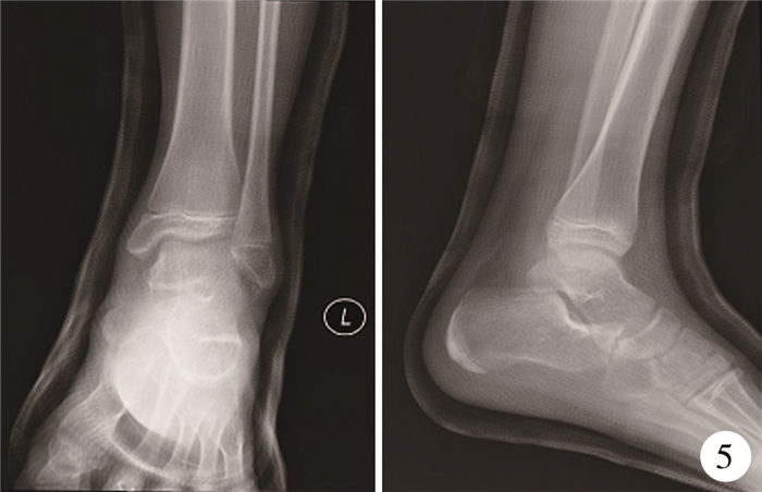
石膏固定4周后,复查X线示距跟、距舟关系良好(图 5),去除石膏,踝关节外观正常,各关节活动可,无疼痛(图 2C、图 2D)。嘱其开始功能锻炼,免负重行走,2周后正常行走。伤后2年6个月,电话随访示左踝关节外观正常,踝关节功能良好,行走无疼痛。

|
图 5 距下关节脱位患者复位4周后左踝正侧位X线片 Fig.5 Radiography of left ankle of a child at 4 weeks after reduction in anterior-posterior and lateral views |
讨论 儿童STJD十分罕见,相关文献较少,均为病例报道[3-8]。STJD根据跟骨前足复合体与距骨的关系可分为内侧脱位、外侧脱位以及前、后脱位4类,其中约80%为内侧脱位。内侧型脱位多因足呈跖屈后暴力内翻,距跟、距舟韧带撕裂所致,外侧型脱位则由外翻暴力引起前后型脱位,外侧型脱位更为少见[7-24]。本文病例年龄为12岁,与既往报道病例相比年龄最小。患者体育锻炼时做侧弓步蹲动作,左踝关节过度内翻造成脱位,属于STJD内侧脱位。
由于STJD罕见,可能造成漏诊或误诊。在诊断过程中需要结合踝关节X线和CT来诊断。X线平片显示为整个足处于内翻位,距下、距舟关节紊乱,但是由于多骨重叠明显,没有特征性的诊断标准,可能造成诊断不清。三维CT可以从多个方向显示畸形,图像清晰。本例诊断过程中,采用双侧对比,发现在横断面上,正常距跟关节之间的跗骨窦呈近似“O形”,而STJD中这一“O形”明显消失,变成不规则形,表明跟骨相对于距骨向内侧脱位;在冠状面和矢状面上也可以看到“O形”,由于本例属于内侧脱位,变形程度不如横断面明显。复位后的CT显示“O形”征恢复正常。因此,我们认为这一征象可以作为STJD的CT诊断标准之一。
对于单纯闭合性STJD,提倡尽早闭合复位,复位前仔细分析移位方向,利用与创伤能量相反的作用机制进行,但禁止反复暴力手法复位,必要时可在麻醉下进行[10, 11, 14]。复位后石膏固定3~4周,如合并骨折需固定5~6周,推荐早期功能康复[19]。尽管大部分闭合性STJD比较容易复位,但文献报道仍有约20%的复位失败率[1]。闭合复位失败的STJD,多因合并距骨、舟骨骨折,伸肌支持带、胫后肌腱、趾短伸肌或者舟骨的阻挡[6, 11, 13, 21]。对于闭合复位失败、开放损伤以及并发其他骨折均应行切开复位,视情况选用内固定或者外固定[7, 9, 17, 18]。Hsu[25]认为对于骨折移位>2 mm、4周内新鲜骨折、骨折块较大外,其他禁忌均应行切开复位。
STJD的短期并发症包括韧带、肌肉及神经损伤、感染,长期并发症包括距下关节、距舟关节骨性关节炎、骨质疏松、距骨缺血性坏死[7, 9, 11, 13-15, 17, 19, 20]。单纯性STJD及早闭合复位多数恢复良好,无骨性关节炎等表现[10, 11, 14]。有研究表明内侧型STJD预后优于外侧型STJD[19],开放性损伤或合并距骨骨折更容易出现距骨缺血性坏死及骨性关节炎,踝关节功能长期随访往往较差[16]。
儿童STJD发病率较低,导致很多临床医生对其认识不足,对于诊断不明确的病例可行CT检查,看是否合并其他骨折或者损伤,复位后还可行MR检查明确软组织及韧带损伤情况[8]。STJD预后多良好,闭合复位简单有效,值得推广。
| [1] |
Perugia D, Basile A, Massoni C, et al. Conservative treatment of subtalar dislocations[J]. Int Orthop, 2002, 26(1): 56-60. DOI:10.1007/s002640100296 |
| [2] |
De Lee JC, Curts R. Subtalar dislocations of the foot[J]. J Bone Joint Surg Am, 1982, 64(3): 433-437. |
| [3] |
Kinik H, Oktay O, Arikan M. Medial subtalar dislocation[J]. Int Orthop, 1999, 23(6): 366-367. DOI:10.1007/s002640050396 |
| [4] |
Garofalo R, Moretti B, Ortolano V, et al. Peritalar dislocations a retrospective study of 18 cases[J]. J Foot Ankle Surg, 2004, 43(3): 166-172. DOI:10.1053/j.jfas.2004.03.008 |
| [5] |
Liu Z, Zhao Q, Zhang L. Medial subtalar dislocation associated with fracture of the posterior process of the talus[J]. J Pediatr Orthop B, 2012, 21(5): 439-442. DOI:10.1097BPB.0b013e328351419c.
|
| [6] |
Hoexum F. Subtalar dislocation: two cases requiring surgery and a literature review of the last 25 years[J]. Arch Orthop Trauma Surg, 2014, 134(9): 1237-1249. DOI:10.1007/s00402-014-2040-6 |
| [7] |
McKeag P, Lyske J, Reaney J, et al. Subtalar dislocation secondary to a low energy injury[J]. BMJ Case Rep, 2015, 2015: bcr2014208828. DOI:10.1136bcr-2014-208828.
|
| [8] |
Gérard R, Kerfant N, Dubois de Mont Marin G, et al. Hawkins' type-Ⅱ talar fracture with subtalar dislocation: A very unusual combination[J]. Orthop Traumatol Surg Res, 2017, 103(3): 403-406. DOI:10.1016/j.otsr.2016.12.009 |
| [9] |
Ramzan MM, Fadl SA. Core curriculum case illustration: medial peritalar dislocation[J]. Emerg Radiol, 2018, 25(3): 329-330. DOI:10.1007/s10140-017-1499-1 |
| [10] |
Benabbouha A. Rare case of pure medial subtalar dislocation in a basketball player[J]. Pan Afr Med J, 2016, 23: 106. DOI:10.11604/pamj.2016.23.106.8848 |
| [11] |
Giannoulis D, Papadopoulos DV, Lykissas MG, et al. Subtalar dislocation without associated fractures: Case report and review of literature[J]. World J Orthop, 2015, 6(3): 374-379. DOI:10.5312/wjo.v6.i3.374 |
| [12] |
DePasse JM, Fantry AJ. Subtle anterior subtalar dislocation[J]. Am J Emerg Med, 2015, 33(10): 1538.e5-e7. DOI:10.1016/j.ajem.2015.07.070 |
| [13] |
Park CH. Fracture of the Posterior Process of the Talus With Concomitant Subtalar Dislocation[J]. J Foot Ankle Surg, 2016, 55(1): 193-197. DOI:10.1053/j.jfas.2015.05.006 |
| [14] |
Azarkane M, Boussakri H, Alayyoubi A, et al. Closed medial total subtalar joint dislocation without ankle fracture: a case report[J]. J Med Case Rep, 2014, 8: 313. DOI:10.1186/1752-1947-8-313 |
| [15] |
Zaraa M, Jerbi I, Mahjoub S, et al. Irreducible subtalar dislocation caused by sustentaculum tali incarceration[J]. J Orthop Case Rep, 2017, 7(1): 58-60. DOI:10.13107/jocr.2250-0685.688 |
| [16] |
Banerjee S, Abousayed MM, Vanderbrook DJ. Lateral subtalar dislocation with tarsometatarsal dislocation: a case report of a rare injury[J]. Case Rep Orthop, 2017, 8090721. DOI:10.1155/2017/8090721 |
| [17] |
Teo AQA, Han F, Chee YH. Unstable open posterior subtalar dislocation treated with a ring external fixator: a case report and review of the literature[J]. J Foot Ankle Surg, 2017, 56(6): 1279-1283. DOI:10.1053/j.jfas.2017.04.032 |
| [18] |
Veltman ES, Steller EJ, Wittich P. Lateral subtalar dislocation: Case report and review of the literature[J]. World J Orthop, 2016, 7(9): 623-627. DOI:10.5312/wjo.v7.i9.623 |
| [19] |
Tanwar YS, Singh S, Arya RK, et al. A closed lateral subtalar dislocation with checkrein deformity of great toe due to entrapment of flexor hallucis longus: a case report[J]. Foot Ankle Spec, 2016, 9(5): 461-464. DOI:10.1177/1938640016630060 |
| [20] |
Harris AP, Ramirez JM, Johnson J. Lateral subtalar fracture-dislocation with maintenance of the talonavicular joint: case study, diagnosis and management[J]. Am J Emerg Med, 2016, 34(10): 2055.e3-e5. DOI:10.1016/j.ajem.2016.03.006 |
| [21] |
Colegate-Stone TJ, James SE. Beware the emergency ankle fracture referral: an unusual case of lateral subtalar joint dislocation secondary to calcaneal fracture with associated lateral malleolus fracture[J]. J Orthop Case Rep, 2015, 5(1): 5-7. DOI:10.13107/jocr.2250-0685.242 |
| [22] |
Ghani Y, Marenah K. Isolated proximal rupture of flexor digitorum longus tendon in a traumatic open subtalar dislocation[J]. Ann R Coll Surg Engl, 2014, 96(6): e10-e12. DOI:10.1308/003588414X13946184902802 |
| [23] |
Gaba S, Kumar A, Trikha V, et al. Posterior dislocation of subtalar joint without associated fracture: a case report and review of literature[J]. J Clin Diagn Res, 2017, 11(9): RD01-RD02. DOI:10.7860/JCDR/2017/27794.10553 |
| [24] |
Hui SH, Lui TH. Anterior subtalar dislocation with comminuted fracture of the anterior calcaneal process[J]. BMJ Case Rep, 2016, 2016: bcr2015213835. DOI:10.1136/bcr-2015-213835 |
| [25] |
Hsu AR, Scolaro JA. Posteromedial approach for open reductionand internal fixation of talar process fractures[J]. Foot Ankle Int, 2016, 37(4): 446-452. DOI:10.1177/1071100716635813 |
 2021, Vol. 20


