早发性脊柱侧弯(early onset scoliosis,EOS)按病因可分为先天性脊柱侧弯、特发性脊柱侧弯、神经肌肉性脊柱侧弯及综合征性脊柱侧弯。其中先天性脊柱侧弯和特发性脊柱侧弯这两类患者肋椎角差和主弯Cobb角均超过20°,被认为具有进展性[1-3]。进展型EOS容易引起严重畸形,其有效治疗是脊柱外科医生的挑战。治疗的目标是控制畸形进展,保持生长发育,待患者达到一定的年龄及体重时再行手术治疗,甚至避免手术。
早发性脊柱侧弯的治疗方法包括随访观察、非手术治疗和手术治疗。其中手术治疗包括半椎体切除和撑开技术,撑开技术包括生长棒(growing rods,GR),钛肋撑开技术(vertical expandable prosthetic titanium rib,VEPTR)和Shilla技术等,手术主要用于治疗中重度早发性脊柱侧弯。
系列石膏作为早发性脊柱侧弯的非手术治疗方法代表,疗效已经得到肯定,但考虑石膏固定脊柱后可能造成肺通气受限,若患有肺部疾病,石膏固定可能会引发呼吸系统并发症。婴幼儿无法配合肺功能检查,不易发现潜在风险[4-8]。对于轻中度进展型EOS,支具治疗作为保守治疗的另一种方式,可弥补石膏固定的弊端。本研究通过分析支具治疗的效果,为治疗进展型早发性脊柱侧弯矫正畸形提供参考依据。
材料与方法 一、临床资料以战略支援部队特色医学中心脊柱外科2011年6月至2019年2月收治的进展型EOS患者为研究对象。纳入标准:①初次发现侧弯畸形时年龄不超过等于5岁;②病因诊断为先天性脊柱侧弯或特发性脊柱侧弯;③随访时间不少于2年。排除标准:①存在明显营养不良表现;②曾行脊柱畸形矫正手术;③非进展型特发性脊柱侧弯、综合征型脊柱侧弯及神经肌肉型脊柱侧弯。末次随访主弯Cobb角较治疗前改善10°的病例归入有效组,改善不超过10°或加重的病例归入无效组。
二、研究方法患者均经支具治疗至少两年,且至少具有治疗前、初次佩戴支具后、末次随访时(去除支具至少24小时)的临床及影像资料。临床资料包括性别,初次支具治疗时年龄,末次随访时年龄,随访时间。拍摄站立位全脊柱正(侧)位X线片,部分患者矢状位无畸形,随访过程中仅拍摄全脊柱正位片。分别测量冠状位(Cobb法)及矢状位畸形角度及平衡以及胸椎高度(T1椎体上终板至T12椎体下终板距离)及胸廓宽度(两侧肋膈角到胸12椎体中线的垂直距离之和)[9]。既往文献报道胸椎高度及胸廓宽度测量可作为代替EOS患者肺功能检查的有效方式[10]。记录顶椎位置及主弯包含椎体数。
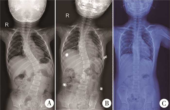
支具治疗方案:支具取模在患者清醒状态下进行,应用点式挤压支具针对全脊柱正侧位X线片,在传统支具原理前提下进行多点挤压。矫正脊柱侧凸畸形的同时,降低因佩戴支具治疗对患者肺功能的影响,提高患者佩戴支具治疗期间的生活质量。初始支具佩戴时间为每天22 h。佩戴支具后每3~6个月随访1次。随访中根据患者侧凸Cobb角、躯干平衡变化情况以及患者的生长发育情况调整支具及佩戴时间。若患者持续6个月侧凸Cobb角及躯干平衡保持稳定,则支具佩戴时间减至每天18 h,若继续保持稳定6个月可改为夜间佩戴,直至停止支具定期观察;若Cobb角加重,则支具佩戴时间保持在每天22 h。典型病例见图 1。

|
图 1 典型病例,患者男性,治疗前年龄31个月,治疗前后及随访拍摄站立位全脊柱正位片,观察畸形变化A:治疗前主弯Cobb角为61°; B:初次矫形后主弯Cobb角为29°; C:佩戴支具6年,末次随访时主弯Cobb角为2° Fig.1 Typical case, male, 31 months old before treatment, Anterior radiographs of the whole spine in standing position before the brace, after the first brace and at last follow-up were taken to observe the deformity changes A:pre-treatment with main Cobb angle of 61 degrees; B:after the first brace with main Cobb angle of 29 degree; C:the main Cobb angle is 2 degrees after 6 years brace treatment |
采用SPSS 20.0进行统计学处理,两组间计量资料对比采用独立样本t检验,两组间计数资料对比采用χ2检验或Fisher精确检验;治疗前后及治疗前与末次随访时影像学参数对比采用配对t检验。P<0.05认为差异有统计学意义。
结果 一、基本情况19例初次支具治疗平均年龄为49.68个月(13~72个月),平均随访时间为49.53个月(24~85个月)。主弯包含椎体数平均为(6.26±1.66)个, 8例顶椎在胸腰段或腰段(47.37%), 3例伴有节段性后凸(15.79%)。至末次随访2例已停止支具治疗,但仍在继续观察随访,避免侧弯再次加重, 17例仍在支具治疗,暂无接受手术患者。其中3例(15.79%)畸形进展大于5°。治疗及随访过程中无并发症发生。
本组患者初次治疗后及末次随访时,主弯Cobb角均较治疗前明显好转(P<0.05)。末次随访时,胸椎后凸角度较治疗前明显增加(P<0.05),胸廓宽度较治疗前明显增加(P<0.05)。但与治疗前相比,初次治疗后胸廓宽度有所下降(P<0.05),胸椎高度在末次随访时及初次佩戴支具时均明显增加(P<0.05)。见表 1。
| 表 1 支具治疗前、初次治疗后与末次随访的影像学表现对比(x±s) Table 1 The radiological change between pretreatment, first in brace and latest follow up(x±s) |
|
|
有效组和无效组性别、初次治疗年龄、末次随访时年龄、支具治疗时间、主弯Cobb角、胸椎后凸、腰椎前凸、胸廓宽度、胸椎高度主弯包含椎体数均无显著差异(P>0.05)。初次治疗后两组间主弯矫正率差异无统计学意义(P>0.05),末次随访时差异具有统计学意义(P<0.05)。病因诊断为特发性脊柱侧弯患者较先天性脊柱侧弯患者矫正效果更好(P<0.05),顶椎位于胸腰段或胸段患者矫形效果更好(P<0.05)。见表 2。
| 表 2 有效组和无效组影像及一般资料对比 Table 2 Radiographic and Treatment Parameters |
|
|
本研究以冠状位主弯矫正度数来判断矫形是否有效。矫正度数超过10°为有效,未超过10°为无效,末次随访时主弯进展大于5°为畸形进展。结果显示病因诊断为特发性脊柱侧弯,顶椎位于胸腰段或腰段患者矫形效果相对良好。有两例患者已得到满意矫形,分别停止支具治疗1年和2年,但仍在继续密切随访观察,直至骨骼发育成熟。3例先天性脊柱侧弯畸形进展大于5°,顶椎均位于胸段,其中两例9岁患者度数大于60°,建议行手术治疗。Bess等[11]报道年龄每增加1岁麻醉诱导出现并发症的风险降低13%,本研究中患者已行支具治疗66.7个月,可安全行手术治疗。
由于本组患者年龄较小,未行肺功能检查。Glotzbecker, M[10]和Johnston CE等[12]报道胸椎高度和胸廓宽度测量可用来评估第一秒用力呼气容积及用力肺活量。本研究中胸椎高度在初次支具治疗及末次随访时均明显增加,胸廓宽度在初次治疗时有所降低,但在末次随访时显著增加,同时末次随访时胸椎后凸也明显增加,表明支具治疗并未影响患者胸廓发育。
目前尚少有支具治疗进展型早发性脊柱侧弯的相关报道。但已有报道表明初次石膏治疗时的年龄、病因学诊断对治疗效果有影响[6, 7]。Mehta[2]发现年龄越小,畸形程度越低(平均32°),石膏治疗效果越好。本研究中两组病例年龄和畸形角度无显著差异。Sanders等[13]报道治疗前主弯Cobb角大于60°治疗后或畸形进展或末次随访大于40°。本组病例主弯Cobb角均不超过60°,否则石膏或手术治疗作为首选。Iorio等[14]发现矫形效果差别在长期随访中更明显,初次石膏治疗时获得的矫正率不能代表石膏治疗的有效性。本研究中所有患者均随访至少两年(24~85个月)。我们发现了同样的现象,初次支具治疗时两组矫形效果无显著差异(P>0.05),但末次随访时(平均随访时间49.73个月)两组间差异具有统计学意义(P<0.05),代偿弯矫正也存在这种现象。
反复麻醉可能导致语言及抽象推理能力发育障碍[15], 近年来美国食品及药物管理局提示部分麻醉药物可能导致大脑发育障碍[16]。支具治疗的优点在于患者不需要接受反复麻醉,可在清醒状态下制作及佩戴。同时避免麻醉状态下的脊髓牵拉损伤。另外支具治疗可及时反馈受压情况,避免皮肤压伤。但由于本研究为一回顾性研究,无法记录患者佩戴支具的依从性。此外,本研究的局限性还包括研究对象年龄较小,未行健康相关的生活质量评估,侧重于影像学评估,以及样本量较小。
综上所述,特发性EOS及顶椎位于胸腰段或腰段支具治疗效果较好。初次支具治疗可获得大部分矫形,这有利于鼓励患者家长监督患者支具佩戴情况。对于那些不能达到矫形的先天性EOS患者,支具治疗至少可推迟手术时间。另外,在治疗及随访过程中,未发现任何支具相关并发症。我们认为,通过规范的支具治疗,主弯Cobb角不超过60°的进展型EOS可取得满意矫形效果。
| [1] |
Fernandes P, Weinstein SL. Natural history of early onset scoliosis[J]. J Bone Joint Surg Am, 2007, 89 Suppl(1): 21-33. DOI:10.2106/JBJS.F.00754 |
| [2] |
Mehta MH. Growth as a corrective force in the early treatment of progressive infantile scoliosis[J]. J Bone Joint Surg Br, 2005, 87(9): 1237-1247. DOI:10.1302/0301-620X.87B9.16124 |
| [3] |
Mehta MH. The rib-vertebra angle in the early diagnosis between resolving and progressive infantile scoliosis[J]. J Bone Joint Surg Br, 1972, 54(2): 230-243. DOI:10.1302/0301-620x.54b2.230 |
| [4] |
郭旭朝, 孟钊, 王飞, 等. 石膏矫形治疗幼儿特发性脊柱侧弯的临床研究[J]. 临床小儿外科杂志, 2020, 19(2): 130-134. Guo XC, Meng Z, Wang F, et al. Clinical study of plaster orthopedic treatment for idiopathic scoliosis toddlers[J]. J Clin Ped Sur, 2020, 19(2): 130-134. DOI:10.3969/j.issn.1671-6353.2020.02.008 (in Chinese) |
| [5] |
李海侠, 吴继功, 宋立志, 等. 系列石膏及支具治疗进展型早发性脊柱侧凸的疗效分析[J]. 中华骨科杂志, 2019, 39(18): 1108-1116. Li HX, Wu JG, Song LZ, et al. The effectiveness analysis of serial cast and bracing for progressive early? onset scoliosis[J]. Chinese Journal of Orthopaedics, 2019, 39(18): 1108-1116. DOI:10.3760/cma.j.issn.0253-2352.2019.18.002 (in Chinese) |
| [6] |
Demirkiran HG, Bekmez S, Celilov R, et al. Serial derotational casting in congenital scoliosis as a time-buying strategy[J]. J Pediatr Orthop, 2015, 35(1): 43-49. DOI:10.1097/BPO.0000000000000229 |
| [7] |
Waldron SR, Poe-Kochert C, Son-Hing JP, et al. Early onset scoliosis:the value of serial risser casts[J]. J Pediatr Orthop, 2013, 33(8): 775-780. DOI:10.1097/BPO.0000000000000072 |
| [8] |
Dhawale AA, Shah SA, Reichard S, et al. Casting for infantile scoliosis:the pitfall of increased peak inspiratory pressure[J]. J Pediatr Orthop, 2013, 33(1): 63-67. DOI:10.1097/BPO.0b013e318264936f |
| [9] |
Gold M, Dombek M, Miller PE, et al. Prediction of thoracic dimensions and spine length on the basis of individual pelvic dimensions:validation of the use of pelvic inlet width obtained by radiographs compared with computed tomography[J]. Spine (Phila Pa 1976), 2014, 39(1): 74-80. DOI:10.1097/BRS.0000000000000036 |
| [10] |
Glotzbecker M, Johnston C, Miller P, et al. Is there a relationship between thoracic dimensions and pulmonary function in early-onset scoliosis[J]. Spine (Phila Pa 1976), 2014, 39(19): 1590-1595. DOI:10.1097/BRS.0000000000000449 |
| [11] |
Bess S, Akbarnia BA, Thompson GH, et al. Complications of growing-rod treatment for early-onset scoliosis:analysis of one hundred and forty patients[J]. J Bone Joint Surg Am, 2010, 92(15): 2533-2543. DOI:10.2106/JBJS.I.01471 |
| [12] |
Johnston CE, McClung A, Fallatah S. Computed Tomography Lung Volume Changes After Surgical Treatment for Early-Onset Scoliosis[J]. Spine Deform, 2014, 2(6): 460-466. DOI:10.1016/j.jspd.2014.04.005 |
| [13] |
Sanders JO, D'Astous J, Fitzgerald M, et al. Derotational casting for progressive infantile scoliosis[J]. J Pediatr Orthop, 2009, 29(6): 581-587. DOI:10.1097/BPO.0b013e3181b2f8df |
| [14] |
Iorio J, Orlando G, Diefenbach C, et al. Serial Casting for Infantile Idiopathic Scoliosis:Radiographic Outcomes and Factors Associated With Response to Treatment[J]. J Pediatr Orthop, 2017, 37(5): 311-316. DOI:10.1097/BPO.0000000000000654 |
| [15] |
Ing C, DiMaggio C, Whitehouse A, et al. Long-term differences in language and cognitive function after childhood exposure to anesthesia[J]. Pediatrics, 2012, 130(3): e476-e485. DOI:10.1542/peds.2011-3822 |
| [16] |
Olutoye OA, Baker BW, Belfort MA, et al. Food and Drug Administration warning on anesthesia and brain development:implications for obstetric and fetal surgery[J]. Am J Obstet Gynecol, 2017, 218(1): 905-907. DOI:10.1016/j.ajog.2017.08.107 |
 2021, Vol. 20


