2. 中国医科大学附属盛京医院小儿泌尿外科(辽宁省沈阳市,110000)
阴囊内脾性腺融合是一种罕见的泌尿生殖系统畸形,由Bostroem[1]于1883年首次报道。该病多见于左侧阴囊,极少见于右侧[2],尚无双侧同时发病的报道。据不完全统计超过70%的病例发病年龄低于20岁,约50%的病例发病年龄低于10岁[3]。男女比例约为16 : 1[4]。由于阴囊内脾性腺融合多为良性,所以儿童阴囊肿物的鉴别诊断对避免不必要的睾丸切除具有重要意义。现将中国医科大学附属盛京医院小儿泌尿外科收治的1例阴囊内脾性腺融合病例报告如下:
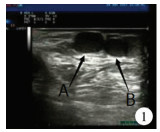
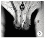
患儿男,13月龄,孕足月剖宫产第一胎,产前检查未见明显异常,无产伤史及出生后缺氧窒息史。因发现左侧阴囊内软质无痛性包块1年而就诊。专科检查:双侧阴囊不对称,左阴囊略增大,内可触及一韧质实性包块,大小约2 cm×1 cm×1 cm,表面皮肤正常,透光试验阴性,无触痛,位于左侧睾丸上方。右侧睾丸及阴茎发育、AFP水平均正常。睾丸附睾精索三维超声:左睾丸上方见1.4 cm×0.8 cm、1.0 cm×0.7 cm包块(图 1),边界清,内呈低回声,CDFI(Color Doppler Flow Imaging,彩色多普勒血流成像)可检出血流信号。超声提示左阴囊内包块。睾丸磁共振平扫:左睾丸上方见两处长T1、稍长T2信号包块,抑脂序列呈高信号。边界清,左侧睾丸受压,睾丸内未见明显异常信号影(图 2)。
|
Download:
|
| 图 1 阴囊异位脾患儿超声结果可见左睾丸上方两包块A:1.4 cm×0.8 cm、B:1.0 cm×0.7 cm,边界清,内呈低回声 Fig. 1 Two masses above left testis were visualized on ultrasonic results in a child of ectopic splenic tissue in scrotum A:1.4*0.8 cm and B:1.0*0.7 cm with a distinct border and lower echoes. | |
|
Download:
|
| 图 2 阴囊异位脾患儿MRIT2抑脂序列可见左侧阴囊内两个包块。边界清,左侧睾丸受压 Fig. 2 Two masses were found in left scrotum on serial lipid-suppressing MRI-T2 in a child of ectopic splenic tissue in scrotum.With a distinct border, left scrotum became compressed | |
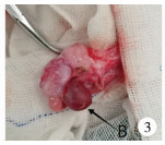
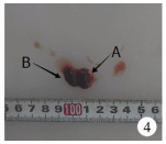
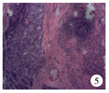
患儿于全麻下行左阴囊肿物探查切除术。术中见左睾丸附睾头处及其近端精索区实性肿物2个(见图 4中A、B箭头所指处),外观呈肉红色,质软,包膜完整,肿物间无相连。精索肿物A约1.5 cm×1.0 cm,睾丸旁肿物B约0.8 cm×0.5 cm, 与睾丸实质界限清(图 3、图 4)。完整切除肿物后即刻送冰冻病理检查,结果为淋巴组织增生,良性,遂保留睾丸。术后第3天复查睾丸精索三维超声提示双侧睾丸及附睾区域未见异常占位病变,常规病理诊断结果为异位脾(图 5)。
|
Download:
|
| 图 3 术中所见睾丸旁肿物,呈深红色,边界清楚 Fig. 3 Intraoperative finding of a dark red mass with a distinct border | |
|
Download:
|
| 图 4 病理大体图片。箭头A所指为睾丸旁精索肿物;箭头B所指为睾丸附睾头处肿物 Fig. 4 Gross pathological gvaph.Arrow A hints at spermatic cord mass off testis while arrow B indicates epididymal mass | |
|
Download:
|
| 图 5 病理镜检图片 肿物A、B中均可见脾组织,肿物B边缘可见少许睾丸曲细精管 Fig. 5 Microscopic examination of pathological specimens Splenic tissue was present in mass A/B.Some twisting ducts lied at the margin of mass B | |
阴囊内脾性腺融合也称阴囊内异位脾、阴囊内副脾。副脾大多数位于脾蒂或胰尾处,也可出现在脾周韧带、大网膜及盆腔内,甚至可以出现在胰腺内。阴囊内脾性腺融合在临床上较为罕见,主要分为连续型和非连续型2种[5]:连续型阴囊内脾性腺融合是指阴囊内异位脾与腹腔内正常脾组织间有脾索相连,经腹股沟管内环穿过腹膜,进入腹腔。此型患儿脾索通常由单一或包含部分脾组织的纤维组织构成,多伴有腹股沟疝和睾丸高位[6-7]。非连续型阴囊内脾性腺融合是指阴囊内异位脾与正常脾组织间无脾索相连,仅有独立包膜与睾丸相间隔。迄今为止,此型未见伴发其他器官畸形的病例报告[8]。本例为非连续型病例。
阴囊内脾性腺融合的发生机制尚不清楚,关于本病的胚胎起源存在两种观点[9]。Sneath[10]认为,覆盖在生殖嵴和脾原基上的腹膜如果出现轻微炎症,可能导致这两个器官发生粘连或部分融合。该理论可以解释脾脏融合现象仅发生于性腺至中肾左部区域这一现象。在连续型脾性腺融合现象中可以观察到,由于性腺的迁移,腹膜粘连会延长,并发展成腹膜内纤维条索(即脾索)。而非连续型脾性腺融合是由于迁移过程中张力过高导致脾索变薄,进而出现自发断裂。Von[11]认为脾原基细胞是通过腹膜后途径与原始性腺相融合。本质上,脾原基前内侧区域与尾生殖褶末端于左胸腹膜内侧相连,生殖褶的末端错位或移位可能导致二者于腹膜后发生融合。该理论可解释右侧非连续型脾性腺融合这一现象。
Curtis等[12-14]认为,副脾组织与正常脾组织的生理学功能相同,可与正常脾组织同时发生病理改变。这一观点被2例阴囊内脾性腺融合患者证实,2例均在疟疾发病时伴阴囊肿胀和疼痛[15]。因此,一些必须行脾切除的血液疾病(如溶血性黄疸、原发性血小板减少性紫癜等)在手术过程中应将所有副脾一并切除。
近半数已报道的脾性腺融合病例发生在儿童群体[9]。Brasch[2]发现行睾丸固定术或睾丸修补术的患者中超过25%存在脾性腺融合,而误将阴囊内脾性腺融合诊断为肿瘤是行根治性睾丸切除术的主要原因。因此,为避免不必要的根治性睾丸切除术,术中对异位脾的组织学鉴定是非常重要的。以下临床要点可能有助于鉴别诊断:①阴囊内异位脾通常为无痛性阴囊肿物,多见于左侧,质软,无活动,透光实验(-),可伴同侧腹股沟疝及睾丸下降不全;②术中可见肿物颜色深红,具有完整包膜,与睾丸或附睾相间隔[9, 16-20]。
随着影像学技术的发展,超声、CT、MRI等检查对本病的诊断有一定的指导意义。超声通常提示肿块信号比睾丸实质略强,可包含多个小的低回声结节,此低回声结节可能与低龄患儿淋巴系统及脾白髓发育高峰有关,因此对本病有较高的诊断价值[21]。但本例患儿年龄较小,肿物发现较早,故未出现此特殊表现。CDFI检查可见肿块内血管密度高[22-25],在连续型脾性腺融合中,超声可发现存在血流信号的条索状组织与肿物相连并进入腹腔[26]。Varma等[27]发现在非连续型脾性腺融合中,肿物与睾丸、附睾间存在分隔,且不依赖睾丸血管进行供血。CT及MRI对连续型脾性腺融合具有较高的诊断价值,通常可见肿物由条索状结构连接进入腹腔。但非连续型脾性腺融合中尚未见到特征性表现[5, 28, 29]。目前相对可靠的术前检查为锝99 m硫胶体显像[30],但明确诊断仍需依赖组织学检查。
该病理论上不需治疗,但多数情况下术前诊断不明确,故需通过手术探查病变性质。因此,怀疑此病时可行手术探查并将肿物切除,但是否需同期行根治性睾丸切除应慎重考虑。
| 1 |
Carragher AM. One hundred years of splenogonadal fusion[J]. Urology, 1990, 35(6): 471-475. DOI:10.1016/0090-4295(90)80097-7. |
| 2 |
Brasch J, Roscher AA. Unusual presentation on the right side of ectopic testicular spleen[J]. Int Surg, 1987, 72(4): 233-234. |
| 3 |
Shen XC, Du CJ, Chen JM, et al. Splenogonadal fusion[J]. Chin Med J (Engl), 2008, 121(4): 383-384. DOI:10.1097/00029330-200802020-00019. |
| 4 |
Pomara G. Splenogonadal fusion:a rare extratesticular scrotal mass[J]. Radiographics, 2004, 24(2): 417. DOI:10.1148/rg.242035171. |
| 5 |
Liu W, Wu R, Guo Z. The diagnosis and management of continuous splenogonadal fusion in a 6-year-old boy[J]. Int Urol Nephrol, 2013, 45(1): 21-4. DOI:10.1007/s11255-012-0349-z. |
| 6 |
Von Hochstetter A. Spleen tissue in the left ovary of the left individual part of a human thoracopagus[J]. Virchows Arch Pathol Anat Physiol Klin Med, 1953, 324(1): 36-54. DOI:10.1007/BF00948094. |
| 7 |
Keane JP, Keane PF. Splenogonadal fusion:an interesting differential for a testicular lump[J]. Ir J Med Sci, 2010, 179(3): 471-472. DOI:10.1007/s11845-010-0489-z. |
| 8 |
Ando S, Shimazui T, Hattori K, et al. Splenogonadal fusion:case report and review of published works[J]. Int J Urol, 2006, 13(12): 1539-1541. DOI:10.1111/j.1442-2042.2006.01598.x. |
| 9 |
May JE, Bourne CW. Ectopic spleen in the scrotum:report of 2 cases[J]. J Urol, 1974, 111(1): 120-123. DOI:10.1016/S0022-5347(17)59904-8. |
| 10 |
Sneath WA. An apparent third testicle consisting of a scrotal spleen[J]. J Anat Physiol, 1913, 47: 340-342. |
| 11 |
Putschar WG, Manion WC. Splenicgonadal fusion[J]. Am J Pathol, 1956, 32(1): 15-33. |
| 12 |
Curtis GM, Movitz D. Surgical Significance of the Accessory Spleen[J]. Ann.Surg, 1946, 123(2): 276-298. DOI:10.1097/00000658-194602000-00008. |
| 13 |
Grossman SL, Goldberg MM, Hermann HB. A case report of ectopic splenic tissue in the scrotum[J]. J Urol, 1959, 81(2): 294-296. DOI:10.1016/S0022-5347(17)66010-5. |
| 14 |
Daniel DS. An unusual case of ectopic splenic tissue resembling a third testicle[J]. Ann Surg, 1957, 145(6): 960-962. DOI:10.1097/00000658-195706000-00016. |
| 15 |
Talmann JM. Nebenmilzen im nebenhodenund samenstrang[J]. Virchows Arch, 1926, 259: 237. DOI:10.1007/BF01890957. |
| 16 |
Settle EB. The surgical significance of accessory spleens with report of two cases[J]. Am J Surg, 1940, 50: 22-26. DOI:10.1016/S0002-9610(40)90357-9. |
| 17 |
Keizur LW. Accessory spleen in the scrotum:Report of two cases[J]. J Urol, 1952, 68(4): 759-762. DOI:10.1016/S0022-5347(17)68278-8. |
| 18 |
Andrews SE, Etter EF. Report of a case of splenic rest in the scrotum[J]. J Urol, 1946, 55: 545-547. DOI:10.1016/S0022-5347(17)69947-6. |
| 19 |
Li X, Ye J, Jiang G. Sonographic diagnosis of splenogonadal fusion in a 2-year-old boy[J]. J Clin Ultrasound, 2017, 45(3): 179-182. DOI:10.1002/jcu.22392. |
| 20 |
Bennett-Jones MJ, Hill CA. Accessory spleen in the scrotum[J]. Br J Surg, 1952, 40(161): 259-262. DOI:10.1002/(ISSN)1365-2168. |
| 21 |
Tate GW, Goforth JL. Accessory spleen in the scrotum:Report of a case[J]. Texas State J Med, 1949, 45(8): 570-573. |
| 22 |
Netto JM, Pérez LM, Kelly DR, et al. Splenogonadal fusion diagnosed by Doppler ultrasonography[J]. Scientific World Journal, 2004, 4(Suppl 1): 253-257. DOI:10.1100/tsw.2004.73. |
| 23 |
Cochlin DL. Splenic-gonadal fusion-the ultrasound appearances[J]. Clin Radiol, 1992, 45(4): 290-291. |
| 24 |
Ferrón SA, Arce JD. Discontinuous splenogonadal fusion:new sonographic findings[J]. Pediatr Radiol, 2013, 43(12): 1652-1655. DOI:10.1007/s00247-013-2730-1. |
| 25 |
Alkhori NA, Barth RA. Pediatric scrotal ultrasound:review and update[J]. Pediatr Radiol, 2017, 47(9): 1125-1133. DOI:10.1007/s00247-017-3923-9. |
| 26 |
Gouw AS, Elema JD, Bink-Boelkens MT, et al. The spectrum of splenogonadal fusion.Case report and review of 84 reported cases[J]. Eur J Pediatr, 1985, 144(4): 316-323. DOI:10.1007/BF00441771. |
| 27 |
Varma DR, Sirineni GR, Rao MV, et al. Sonographic and CT features of splenogonadal fusion[J]. Pediatr Radiol, 2007, 37(9): 916-919. DOI:10.1007/s00247-007-0526-x. |
| 28 |
Li YH. Preoperative detection of splenogonadal fusion by CT[J]. Surg Radiol Anat, 2009, 31(9): 733-735. DOI:10.1007/s00276-009-0507-x. |
| 29 |
Croxford WC, Pfistermuller KL, Scott F, et al. Splenogonadal fusion presenting clinically and radiologically as a seminoma[J]. Urol Case Rep, 2015, 3(6): 204-205. DOI:10.1016/j.eucr.2015.06.007. |
| 30 |
Steinmetz AP, Rappaport A, Nikolov G, et al. Splenogonadal fusion diagnosed by spleen scintigraphy[J]. J Nucl Med, 1997, 38(7): 1153-1155. |
 2019, Vol. 18


