成骨不全症(osteogenesis imperfecta, OI)即脆骨病,常表现为骨质疏松、骨脆性增加和易骨折。据报道其发病率约1/15 000[1],严重者可出现骨骼畸形甚至围生期死亡,另有部分患儿伴随身材矮小、鸡胸、脊柱侧弯、关节韧带松弛及肌肉薄弱等症状[2]。
目前,OI的治疗方案主要包括药物治疗和髓内钉固定术2种。1959年Sofield和Millar首次报道了多处截骨联合髓内钉固定治疗成骨不全的案例,发现患儿近期治疗效果较为满意。但是随着患儿骨骼的生长发育,需要再次手术更换髓内钉,且术后易发生再骨折、畸形愈合和内固定移位等并发症[3]。为解决上述问题,可延长髓内钉应运而生,它包括近端和远端两部分,置入长骨的骨骺后可随患儿的骨骼生长而延长。1963年,Bailey和Dubow制作了一种可延长的髓内固定装置,降低了再手术的机率[4]。但该内固定方式术后并发症的发生率较高,1986年,英国谢菲尔德儿童医院团队设计的Sheffield Telescopic Intramedullary Rod(SF)改善了BD系统的不足,将T端固定在钉末端,避免了脱落移位,但该系统置入需切开关节[5]。2003年,加拿大Pega医疗公司生产的Fassier-Duval Telescopic IM System(FD),可从骨近端单侧置入,并且在钉两端增加了螺纹以使其固定在骨骺,不需额外切开关节[6]。而Dyna-Locking/Interlocking Telescopic Rod(IL)由韩国Cho TJ等[7]于2007年报道,该装置同样可从近端单侧置入,末端没有螺纹结构及T端,但需加用一枚锁定钉横行穿过钉末端的小洞来固定。
自上述改良内固定装置问世以来,已陆续有相关研究报道它们的安全性,但不同研究间结论并不完全一致。因此,本研究将对收集到的文献中报道的术后并发症及随访结果进行Meta分析。
材料与方法 一、检索策略检索PubMed、EmBase、Cochrane图书馆、万方数据资源系统及中国期刊网全文数据库中使用可延长髓内钉固定治疗儿童成骨不全下肢骨折的研究,检索时限为1986年4月至2018年1月。中文检索词为:可延长髓内钉;固定;儿童成骨不全。英文检索词为:“osteogenesis imperfecta;Bailey-Dubow;Sheffield;Fassier-Duval;Dyna-Locking;telescopic”。为尽可能纳入所有符合标准的研究,通过搜索研究的参考文献部分进一步查找所需文献。当研究内容包含可延长及非延长髓内钉治疗时,仅纳入可延长髓内钉治疗后数据。由2位评估者对文章的全文进行查阅,并独立判断是否纳入,当2位评估者意见不统一时,由较高年资者作出决定。
二、纳入及排除标准 (一) 纳入标准① 1986年4月至2018年1月间使用内固定治疗成骨不全下肢骨折的研究;②研究对象为儿童;③干预措施:使用可延长髓内钉系统(SF、FD、IL),且运用传统手术方法;④有详细的病例信息。
(二) 排除标准① 中文、英文语种以外的文献;②受试者接受非常规手术方法或非初次手术;③无详细病例信息;④无术后并发症记录及随访结果;⑤单个病例报告;⑥术后使用双磷酸盐治疗;⑦非随机研究方法评分(methodological index for non-randomized studies,MINORS) < 12分[8]。
(三) 结局指标① 髓内钉相关并发症(钉弯曲、钉移位、过度延长以及穿破骨膜)发生例数;②骨相关并发症(愈合不佳、再次发生骨折)发生例数;③再手术发生例数。
三、数据提取和质量评价收集的信息主要包括:第一作者、发表年限、样本数量及分布、平均年龄及范围、平均随访时间及范围、内固定方式及安全性随访结果。鉴于本研究纳入的文献多采用非临床随机对照试验设计,质量评价采用非随机研究方法MINORS进行评分(表 1)[8],评价指标共8项,每一项可评0~2分(0分表示未报道;1分表示报道但信息不充分;2分表示报道且提供充分信息)。质量评价由两位评估者独立进行,当评价结果不一致时,参考原文进行讨论直到意见达成一致。
|
|
表 1 MINORS评价条目 Table 1 Evaluation entries of MINORS |
采用Stata10.0进行Meta分析,合并分析结果以发生率及其95%CI表示。依据异质性检验结果分别采用固定或随机效应模型[9, 10]。当研究间异质性I2≤25%时采用固定效应模型,反之则采用随机效应模型[11, 12]。对骨相关并发症、髓内钉相关并发症及再手术发生率进行敏感性分析以探讨某项研究的剔除是否会对合并结果产生影响[13]。发表偏倚结果采用漏斗图、Egger及Begg检验进行分析,当P < 0.05时认为存在发表偏倚。
结 果 一、文献检索结果经筛选过后纳入本次Meta分析的文献共8篇[5, 7, 14-19]。筛选流程见图 1。以8篇文献中报道的258例经SF、FD、IL可延长髓内固定的OI患儿下肢骨为研究对象,其中股骨157例(57.4%),胫骨101例(42.6%)。患儿年龄范围为3.1~8.0岁,均为首次接受手术治疗,术后平均随访17~228个月(表 2)。

|
Download:
|
| 图 1 文献筛选流程图 Fig. 1 Flow chart of literature screening | |
|
|
表 2 纳入文献的基本特征 Table 2 Basic characteristics of each literature entry |
8项研究均对髓内钉相关并发症情况进行了统计。异质性检验结果I2=73.8%,故采用随机效应模型进行合并分析,得出髓内钉相关并发症发生率为25.00%(95% CI:15.00% ~34.00%)(图 2),其中钉移位占13.54%、钉弯曲占6.18%、穿破骨膜3.87%、过度延长占2.5%(表 3);敏感性分析结果见图 3,发现排除J.M.Wilkinson的研究后,研究间异质性显著降低。图 4对髓内钉相关并发症的发表偏倚进行了检测,发现漏斗图中散点分布对称性良好。Egger和Begg检验结果同样显示无发表偏倚(PEgger=0.248,PBegg=0.386)。
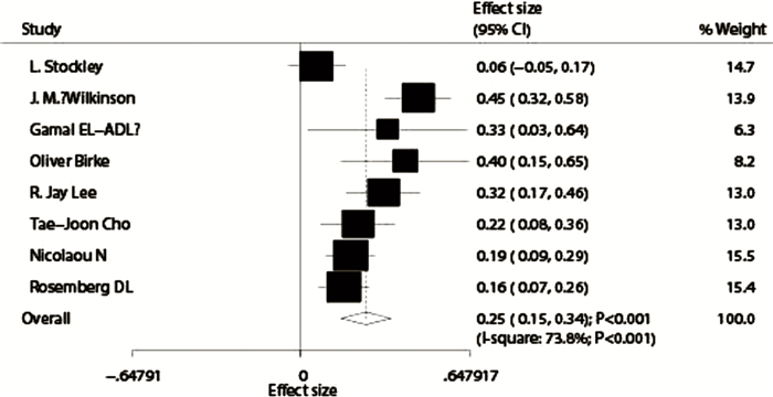
|
Download:
|
| 图 2 髓内钉相关并发症发生率森林图 Fig. 2 Forest plot of the incidence of intramedullary nail-related complications | |
|
|
表 3 术后并发症发生率 Table 3 Postoperative complication rate |
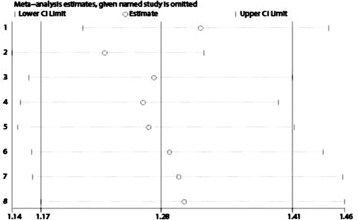
|
Download:
|
| 图 3 髓内钉相关并发症发生率敏感性分析 Fig. 3 Sensitivity analysis of the incidence of intramedullary nail-related complications | |
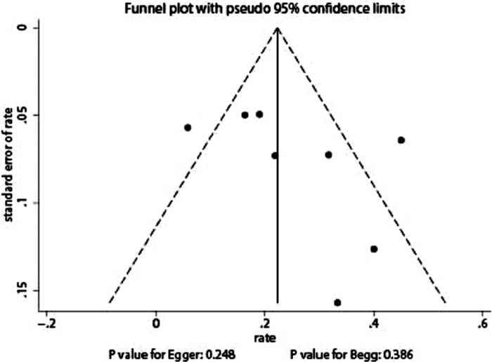
|
Download:
|
| 图 4 髓内钉相关并发症发生率漏斗图 Fig. 4 Funnel plot of the incidence of intramedullary nail-related complications | |
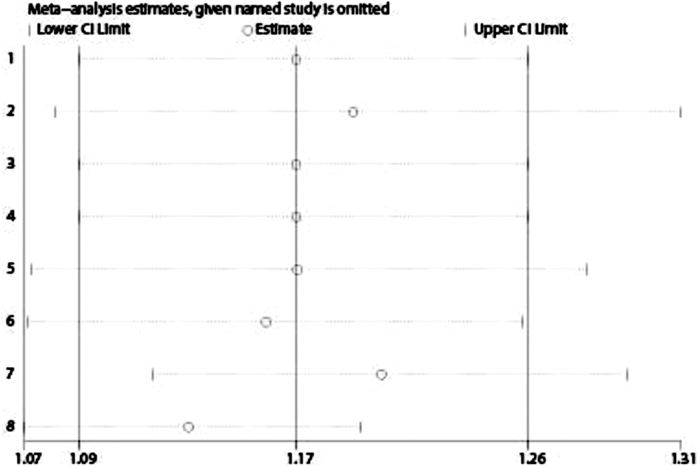
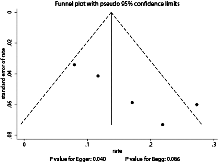
5项研究对骨相关并发症的发生情况进行了统计,剩余3项研究显示发生人数为0,因而不参与合并分析过程。异质性检验结果I2=59.2%,故采用随机效应模型进行合并,得到骨相关并发症发生率为16.00%(95% CI:9.00% ~23.00%)(图 5),其中再次发生骨折占14.66%、愈合不佳占2.93%(表 3)。敏感性分析结果见图 6,排除Rosemberg DL的研究后,研究间异质性显著降低。对骨相关并发症的发表偏倚进行检测,通过漏斗图(图 7)发现对称性一般。Egger和Begg检验结果提示可能存在发表偏倚(PEgger=0.040,PBegg=0.086)。
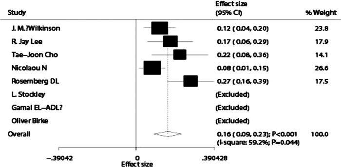
|
Download:
|
| 图 5 骨相关并发症发生率森林图 Fig. 5 Forest plot of the incidence of bone-related complications | |
|
Download:
|
| 图 6 骨相关并发症发生率敏感性分析 Fig. 6 Sensitivity analysis of the incidence of bone-related complications | |
|
Download:
|
| 图 7 骨相关并发症发生率漏斗图 Fig. 7 Funnel plot of bone-related complication rate | |
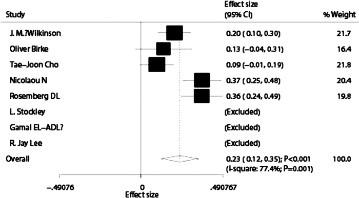
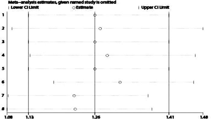
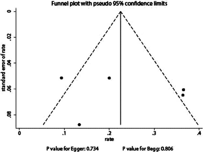
5项研究对再手术的发生率进行了统计,剩余3项研究指出再手术人数为0,因而不参与合并分析的过程。异质性检验结果I2=77.4%,故采用随机效应模型进行合并分析,发现再手术发生率为23.00%(95% CI:12.00% ~35.00%)(图 8)。其敏感性分析结果见图 9,排除任意一项研究并不能显著改善异质性。对再次手术发生率的发表偏倚进行检测,漏斗图(图 10)指出散点分布对称性较好。Egger和Begg检验结果显示出无发表偏倚(PEgger=0.734;PBegg=0.806)。
|
Download:
|
| 图 8 再手术率森林图 Fig. 8 Forest plot of reoperation rate | |
|
Download:
|
| 图 9 再手术发生率敏感性分析 Fig. 9 Sensitivity analysis of the incidence of reoperation | |
|
Download:
|
| 图 10 再手术发生率的漏斗图 Fig. 10 Funnel plot of reoperation rate | |
因废用性骨质疏松和局部畸形,儿童成骨不全症常伴发骨折,以下肢骨折最为多见,单凭夹板或骨牵引等非手术疗法很难在治疗骨折的同时避免畸形愈合及再骨折[20]。因OI患儿骨质较脆,外固定支架或钢板容易导致新发骨折和畸形加重,故不建议采用[21]。目前,对于OI患儿再发骨折、进行性畸形及功能受损,严格按照指征行手术治疗已成为共识[5]。根据骨折和畸形的特点,多段截骨加髓内钉内固定是公认的主流手术方式,该术式不仅可以有效矫正畸形、增强肢体强度、减少再骨折发生率,还可延长肢体[7]。在可延长钉发展成熟以前,医生多使用Kirschner钉、Rush钉、Ender钉和elastic钉,这类非延长髓内钉固定后短期效果均较好,并可起到预防再次骨折的作用[22-24]。但随着患儿骨骼生长速度逐渐加快,当生长板距离钉越来越远后,应力作用容易诱发骨骼畸形及再骨折。BD可延长髓内钉系统被发明后,发达国家便开始较为广泛地使用,但术后存在较高的并发症(包括T端脱落、钉移位、再骨折等)发生率[25-29]。
敏感性分析结果显示J.M.Wilkinson[5]与其他研究间的异质性较大,从该研究报道的时间来看,彼时SF钉系统尚处于使用初期阶段,手术技术尚未完全成熟,导致术后效果欠佳;且该研究纳入的手术例数多、随访时间长,因此可能造成并发症发生率相对其他研究较高的现象。漏斗图分析中提示纳入的研究无显著性偏倚,这进一步说明本次分析的结果具有一定的代表性。
仅5项研究对骨相关并发症的发生情况进行统计,合并后骨相关并发症发生率为16.00%,其中再骨折占比最多(14.66%)。排除Rosemberg DL[19]的研究后,异质性同样显著降低,可能与患儿病情严重程度及不同的OI类型有关;另外该报道发表于2018年,内固定使用FD系统,且随访时间较久,手术技术更加成熟;在漏斗图分析结果中也提示存在发表偏倚,可能与样本量不足有关。
仅5项研究对再手术率进行统计,整合分析后显示数据间仍存在一定异质性,且排除任意一项研究并不能显著改善异质性,这可能与各中心对再手术指征评价标准存在差异、手术医师熟练程度不一及不同年龄患儿对手术接受程度不同有关。将研究合并后发现,因以上并发症需要再次手术的患儿占23.00%,漏斗图显示散点对称性较好,无显著性发表偏倚。2017年Scollan等报道了关于非延长髓内钉治疗OI患儿下肢骨折术后并发症Meta分析[30],其中钉移位发生率为25.7%,骨相关并发症为19.51%,而再手术率达39.39%;尽管术后骨畸形愈合发生率仅4.26%,但可延长钉似乎达到了更好的效果(骨畸形愈合发生率仅2.93%)。虽然使用可延长钉可能造成其特有的并发症,但发生率并不高,加上OI本身的病理特点,手术并不能改变患儿的基因缺陷,因此术后再骨折、畸形愈合在所难免。所以,使用髓内钉治疗的主要目的还是提供更坚固的支撑,降低再骨折的发生,提高OI患儿的生活质量。
在搜索文献过程中,作者也收集到一些随访时间长、术后并发症记录较详细的文献,但是因为患者超龄、非首次手术、上肢骨折手术、未明确记录相应内固定类型以及术后加用双磷酸盐等因素未能纳入本次Meta分析,但其中的结果和结论仍具有一定的参考价值[31-35]。
本研究也存在一定的不足。首先,本次纳入的研究均为回顾性研究,缺乏更加严谨、更具科学性的科研设计模式(但由于外科手术本身限制,所以不能排除选择偏倚的影响);其次,本次搜索文献的语种为英语和汉语,存在一定程度的语种偏移,虽然采取了广泛检索策略,但由于纳入的研究较少,必然存在一定的发表偏倚,需要在以后的研究中扩大样本量;再者,由于外科手术的特殊性,不同的外科医生在手术技巧、术中应对不同情况的处理和术后治疗、护理等方面难以达到统一标准,因此可能存在实施偏倚,所以还需要更详细的数据来进一步证实本次研究的结论。
| 1 |
Forlino A, Marini JC. Osteogenesis imperfecta[J]. Lancet, 2016, 387(10028): 1657-1671. DOI:10.1016/S0140-6736(15)00728-X. |
| 2 |
潘少川. 实用小儿骨科学[M]. 第3版. 人民卫生出版社, 2016: 194-195. Pan SC. Practice of Pediatric Orthopedics[M]. Third Edition: People's Medical Publishing Press, 2016: 194-195. |
| 3 |
Sterian A, Balanescu R, Barbilian A, et al. Early telescopic rod osteosynthesis for Osteogenesis Imperfecta patients[J]. J Med Life, 2015, 8(4): 544-547. |
| 4 |
Lang-Stevenson AI, Sharrard WJ. Intramedullary rodding with Bailey-Dubow extensible rods in osteogenesis imperfecta.An interim report of results and complications[J]. J Bone Joint Surg Br, 1984, 66(2): 227-232. |
| 5 |
Wilkinson JM, Scott BW, Clarke AM, et al. Surgical stabilisation of the lower limb in osteogenesis imperfecta using the Sheffield Telescopic Intramedullary Rod System[J]. J Bone Joint Surg Br, 1998, 80(6): 999-1004. DOI:10.1302/0301-620X.80B6.8667. |
| 6 |
Fassier F. Fassier-Duval Telescopic System:How I Do It?[J]. J Pediatr Orthop, 2017, 37(6): S48-S51. DOI:10.1097/BPO.0000000000001024. |
| 7 |
Cho TJ, Choi IH, Chung CY, et al. Interlocking telescopic rod for patients with osteogenesis imperfecta[J]. J Bone Joint Surg Am, 2007, 89(5): 1028-1035. DOI:10.2106/JBJS.F.00814. |
| 8 |
曾宪涛, 庄丽萍, 杨宗国, 等. Meta分析系列之七:非随机试验性研究、诊断性试验及动物实验的质量评价工具[J]. 中国循证心血管医学杂志, 2012, 4(6): 496-499. DOI:10.3969/j.1674-4055.2012.06.003. Zeng XT, Zhuang LP, Yang ZG, et al. The seventh article of the Meta analysis series:the quality evaluation tool of the non-randomized study, diagnostic study and animal experiment[J]. Chin J Evid Based Cardiovase Med, 2012, 4(6): 496-499. DOI:10.3969/j.1674-4055.2012.06.003. |
| 9 |
DerSimonian R, Laird N. Meta-analysis in clinical trials[J]. Control Clin Trials, 1986, 7: 177-188. DOI:10.1016/0197-2456(86)90046-2. |
| 10 |
Ades AE, Lu G, Higgins JP. The interpretation of random-effects metaanalysis in decision models[J]. Med Decis Making, 2005, 25: 646-654. DOI:10.1177/0272989X05282643. |
| 11 |
Deeks JJ, Higgins JPT, Altman DG.Analyzing data and undertaking meta-analyses.In: Higgins J, Green S, eds.Cochrane Handbook for Systematic Reviews of Interventions 5.0.1.Oxford, UK: The Cochrane Collaboration: 2008;chap 9.
|
| 12 |
Higgins JP, Thompson SG, Deeks JJ, et al. Measuring inconsistency in meta-analyses[J]. BMJ, 2003, 327(7414): 557-560. DOI:10.1136/bmj.327.7414.557. |
| 13 |
Tobias A. Assessing the influence of a single study in meta-analysis[J]. Stata Tech Bull, 1999, 47: 15-17. |
| 14 |
Birke O, Davies N, Latimer M, et al. Experience with the Fassier-Duval telescopic rod:first 24 consecutive cases with a minimum of 1-year follow-up[J]. J Pediatr Orthop, 2011, 31(4): 458-464. DOI:10.1097/BPO.0b013e31821bfb50. |
| 15 |
Lee RJ, Paloski MD, Sponseller PD, et al. Bent Telescopic Rods in Patients With Osteogenesis Imperfecta[J]. J Pediatr Orthop, 2016, 36(6): 656-660. DOI:10.1097/BPO.0000000000000509. |
| 16 |
Stockley I, Bell MJ, Sharrard WJ. The role of expanding intramedullary rods in osteogenesis imperfecta[J]. J Bone Joint Surg Br, 1989, 71(3): 422-427. |
| 17 |
El-Adl G, Khalil MA, Enan A, et al. Telescoping versus non-telescoping rods in the treatment of osteogenesis imperfecta[J]. Acta Orthop Belg, 2009, 75(2): 200-208. |
| 18 |
Nicolaou N, Bowe JD, Wilkinson JM, et al. Use of the Sheffield telescopic intramedullary rod system for the management of osteogenesis imperfecta:clinical outcomes at an average follow-up of nineteen years[J]. J Bone Joint Surg Am, 2011, 93(21): 1994-2000. DOI:10.2106/JBJS.J.01893. |
| 19 |
Rosemberg DL, Goiano EO, Akkari M, et al. Effects of a telescopic intramedullary rod for treating patients with osteogenesis imperfecta of the femur[J]. J Child Orthop, 2018, 12(1): 97-103. DOI:10.1302/1863-2548.12.170009. |
| 20 |
宿玉玺, 赵仁欢, 覃佳强, 等. Rush钉矫治成骨不全儿童股骨畸形的疗效观察[J]. 第三军医大学学报, 2015, 37(3): 238-242. DOI:10.16016/j.1000-5404.201409066. Su YX, Zhao RH, Qin JQ, et al. Clinical efficacy of Rush rods in correction of femoral deformity in children's tosteogenesis imperfecta[J]. J Third Mil Med Univ, 2015, 37(3): 238-242. DOI:10.16016/j.1000-5404.201409066. |
| 21 |
任秀智, 曾裴, 李冰, 等. 多段截骨矫形髓内固定治疗儿童成骨不全症[J]. 中华骨科杂志, 2012, 32(5): 477-481. DOI:10.3760/cma.j.issn.0253-2352.2012.05.018. Ren XZ, Zeng P, Li B, et al. Multi-section osteotomy and intramedullary fixation for children with osteogenesis imperfecta[J]. Chinese Journal of Orthopaedics, 2012, 32(5): 477-481. DOI:10.3760/cma.j.issn.0253-2352.2012.05.018. |
| 22 |
Joseph B, Rebello G, B CK. The choice of intramedullary devices for the femur and the tibia in osteogenesis imperfecta[J]. J Pediatr Orthop B, 2005, 14(5): 311-319. DOI:10.1097/01202412-200509000-00001. |
| 23 |
Mulpuri K, Joseph B. Intramedullary rodding in osteogenesis imperfecta[J]. J Pediatr Orthop, 2000, 20(2): 267-273. |
| 24 |
Boutaud B, Laville JM. Elastic sliding central medullary nailing with osteogenesis imperfecta.Fourteen cases at eight years follow-up[J]. Rev Chir Orthop Reparatrice Appar Mot, 2004, 90(4): 304-311. DOI:10.1016/S0035-1040(04)70125-7. |
| 25 |
Bailey RW, Dubow HI. Studies of longitudinal bone growth resulting in an extensible nail[J]. Surg Forum, 1963, 14: 455-458. |
| 26 |
Luhmann SJ, Sheridan JJ, Capelli AM, et al. Management of lower-extremity deformities in osteogenesis imperfecta with extensible intramedullary rod technique:a 20-year experience[J]. J Pediatr Orthop, 1998, 18(1): 88-94. |
| 27 |
Middleton RW. Closed intramedullary rodding for osteogenesis imperfecta[J]. J Bone Joint Surg Br, 1984, 66(5): 652-655. |
| 28 |
Nicholas RW, James P. Telescoping intramedullary stabilization of the lower extremities for severe osteogenesis imperfecta[J]. J Pediatr Orthop, 1990, 10(2): 219-223. DOI:10.1097/01241398-199003000-00017. |
| 29 |
Bailey RW. Further clinical experience with the extensible nail[J]. Clin Orthop Relat Res, 1981(159): 171-176. |
| 30 |
Scollan JP, Jauregui JJ, Jacobsen CM, et al. The Outcomes of Nonelongating Intramedullary Fixation of the Lower Extremity for Pediatric Osteogenesis Imperfecta Patients:A Meta-analysis[J]. J Pediatr Orthop, 2017, 37(5): e313-e316. DOI:10.1097/BPO.0000000000000970. |
| 31 |
Cho TJ, Kim JB, Lee JW, et al. Fracture in long bones stabilised by telescopic intramedullary rods in patients with osteogenesis imperfecta[J]. J Bone Joint Surg Br, 2011, 93(5): 634-638. DOI:10.1302/0301-620X.93B5.25499. |
| 32 |
Sterian A, Balanescu R, Barbilian A, et al. Osteosynthesis in Osteogenesis Imperfecta, telescopic versus non-telescopic nailing[J]. J Med Life, 2015, 8(4): 563-565. |
| 33 |
Grossman LS, Price AL, Rush ET, et al. Initial Experience With Percutaneous IM Rodding of the Humeri in Children With Osteogenesis Imperfecta[J]. J Pediatr Orthop, 2018, 38(9): 484-489. DOI:10.1097/BPO.0000000000000856. |
| 34 |
Azzam KA, Rush ET, Burke BR, et al. Mid-term Results of Femoral and Tibial Osteotomies and Fassier-Duval Nailing in Children With Osteogenesis Imperfecta[J]. J Pediatr Orthop, 2018, 38(6): 331-336. DOI:10.1097/BPO.0000000000000824. |
| 35 |
Sa-Ngasoongsong P, Saisongcroh T, Angsanuntsukh C, et al. Using humeral nail for surgical reconstruction of femur in adolescents with osteogenesis imperfecta[J]. World J Orthop, 2017, 8(9): 735-740. DOI:10.5312/wjo.v8.i9.735. |
 2019, Vol. 18


