骨折后骨囊肿样改变(Postfracture cyst-like lesions,PCLL)在儿童骨折治疗中报道很少,病因尚不明确,具有自限性,并对骨折塑形没有影响。既往病例报道中主要通过放射线检查偶然发现,且大多来自儿童桡骨远端骨折[1]。本文总结1例儿童胫骨骨折后骨囊肿样改变的临床特点,并对既往相关研究结合本病例的特征进行文献分析。
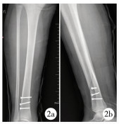
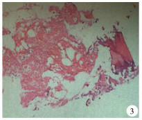
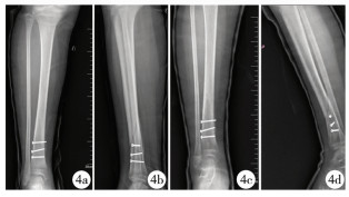
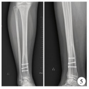
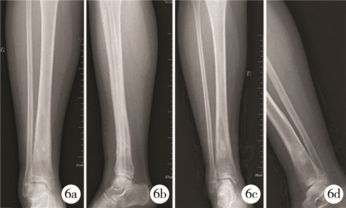
患儿,女,13岁,于2016年7月9日因“不慎自行摔伤2 h”到大连市儿童医院骨科门诊就诊,X线检查显示骨折对位对线尚可(图 1),建议石膏外固定并门诊随诊,1周后X线检查结果提示正位X线上骨折较前稍移位,侧位片未见明显变化,向患儿家长交代病情后,患儿家长为避免骨折移位增加,强烈要求手术并骨折内固定治疗。于2016年7月17日在全麻下行胫骨骨折切开复位拉力螺钉内固定手术并超膝石膏外固定(图 2),术中见骨膜未断裂,未见骨组织病理改变,术后每2周复查1次X线,术后1个月(图 4)发现胫骨原骨折线远端骨皮质变薄,术后3个月PCLL形成(图 4),PCLL有一定自愈性,继续给予石膏外固定,术后3个月拆除石膏负重行走,随诊患儿无疼痛等症状,PCLL范围不断增大,术后1年PCLL未见好转(图 5),且病灶X线下体积达9 cm×3 cm×2 cm,为避免发生病理性骨折,于2017年7月13日在全麻下予拉力螺钉内固定装置取除、病灶刮除、异体骨植骨手术治疗,术中见骨折表面骨皮质变薄,骨刀切除1.5 cm×1.0 cm骨皮质后,见肿物为单房囊性,内容物为血性液体,术后病理检查结果支持囊肿改变,并内含脂肪组织及少量骨组织(图 3),术后随访4个月(图 6),病灶愈合好,并继续随访中。

|
Download:
|
| 图 1 a:右侧胫骨远端外伤后正位X线片可见骨折对位对线尚可;b:右侧胫骨远端外伤后侧位X线片可见骨折对位对线尚可;c:外伤石膏固定1周后正位X线片;d:外伤石膏固定1周后侧位X线片 Fig. 1 a:Radiographs of the right tibia at the time of Injury show the posteroanterior position of fracture can be accepted.b:Radiographs of the right tibia at the time of Injury show the lateral position of fracture can be accepted.c:Posteroanterior Radiographs of the right tibia one week after Injury.d:Lateral Radiographs of the right tibia one week after Injury | |
|
Download:
|
| 图 2 a:第1次手术后1 d正位X线片示骨折对位对线良好,内固定可靠;b:第1次手术后1 d侧位X线片示骨折对位对线良好,内固定可靠 Fig. 2 a:posteroanterior Radiographs of the right tibia at the time of one day after the first operation.b:Lateral Radiographs of the right tibia at the time of one day after the first operation | |
|
Download:
|
| 图 3 病理结果(HE染色放大100) Fig. 3 Pathology Result (HE staining, magnify 100 multiple) | |
|
Download:
|
| 图 4 a、b:术后1个月正位X线片提示骨折线远端骨皮质变薄;c、d:术后3个月PCLL形成 Fig. 4 a、b: Radiographs of the right tibia at the time of one month after the first operation, shows that the distal cortex of the fracture is thinning; c、d: Radiographs of the right tibia at the time of three month after the first operation shows PCLL | |
|
Download:
|
| 图 5 第1次手术术后1年正侧位X线片提示PCLL不断扩大 Fig. 5 Radiographs of the right tibia at the time of one year after the first operation, shows the PCLL is expanding | |
|
Download:
|
| 图 6 a:第2次手术后1个月正位X线片;b:第2次手术后1个月侧位X线片;c:第2次手术术后4个月正位X线片;d:第2次手术术后个4月侧位X线片 Fig. 6 a:posteroanterior Radiographs of the right tibia at the time of one month after the second operation.b:Lateral Radiographs of the right tibia at the time of one month after the second operation.c:posteroanterior Radiographs of the right tibia at the time of three month after the second operation.d:posteroanterior Radiographs of the right tibia at the time of three month after the second operation | |
1969年Levine BS等[2]首次报道13岁男童腓骨骨折后PCLL形成。目前英文文献中共有30余例病例报道,大多为桡骨远端骨折后出现PCLL,都发生在骨折后1~2个月,且病灶明显具有自限性[3]。
PCLL分为两种类型:一种是暂时型,儿童多见,病灶多为圆形或者椭圆形,直径1 cm左右,周围1 cm范围出现骨压缩表现,愈合时间多在1~3个月;另一种是中心膨胀型,儿童和成人均有报道,与暂时型有明显区别,病灶大于1 cm,同样具有自限性,但是病程长,多在1~3年后愈合[1]。
Pfister-Goedeke L[11]认为是骨折后的骨膜过度反应造成了PCLL,而后自行吸收。Philips CD等[12]认为是骨髓腔内出血造成PCLL, 后自行吸收。
Malghem J和Maldague B[5, 6]通过对PCLL患者CT扫描发现了病灶内的脂肪颗粒,并认为脂肪液化是PCLL形成的主要原因,这样也就解释了为什么PCLL在外伤4~8周后出现,而不是在骨折后的早期。考虑原因为血肿吸收或者钙化后才开始脂肪液化。Ball CM等[4]也支持该观点,推测骨折后骨髓损伤使骨髓内脂肪通过破裂的骨皮质转移到骨膜下,骨膜下血肿黏附,血肿吸收后脂肪液化出现PCLL。这就是脂肪内容物学说。
该学说后来又被多位学者完善:认为PCLL内容物多为血肿、脂肪混合物。骨折造成了髓内脂肪颗粒漂浮在出血表面,进入骨折周围骨膜下,骨膜完整,血肿吸收后,脂肪液化侵蚀骨皮质,并向周围扩展,PCLL形成。骨皮质结痂,但是与骨膜分离,使得脂肪液化有了空间,未完全吸收的血肿为PCLL形成提供养料,而后逐渐被骨痂、新生骨充填,最终该部位髓腔重建[7, 8]。
|
|
表 1 相关文献汇总 Table 1 total ralate papers |
儿童骨膜相对成人更加松弛、厚度低,但是更加牢固,容易在骨折后出现保持完整并出现骨膜与长骨骨皮质间的空间,为PCLL提供场所。造成病例多集中在桡骨远端骨折的原因包括:①儿童桡骨远端骨折病例多,出现PCLL几率大;②儿童骨折随访时间多在6周以内,并未随访到病灶出现;③该处骨组织血液供应较差,局部血肿吸收慢,容易与髓内脂肪形成黏附混合物[14-16]。
二、病例少有报道原因分析PCLL罕有报道的主要原因有以下几点:①骨皮质的损伤既要有足够骨髓脂肪外渗,同时骨膜连续性不能受到破坏,从而形成一定的空间使血肿和脂肪积聚;②骨折时出血量较多,为脂肪颗粒转移提供条件;③脂肪液化的偶然发生;④大多数骨折随诊时间4~6周,随访时间不足,未能随访到该病理变化,后PCLL自愈。
三、骨囊肿与PCLL对比骨囊肿是一种充满液体的腔性骨组织良性病变,好发年龄5~15岁,手术治疗具有较高复发率。病因包括:肿瘤学说,炎症学说,骨发育异常学说,静脉淤滞学说等,其中静脉淤滞学说为大多学者推崇。治疗主要目的是防止病理性骨折,促进病变愈合[9, 10]。
PCLL内容物伴有脂肪组织,好发年龄1.5~15岁,病程具有明显的自限性[1]。病因包括:骨内膜过度反应[11],骨髓腔内出血[12],脂肪内容物学说[7]等,目前脂肪内容物学说为大多学者推崇。
四、本例PCLL特点及病因分析本患儿存在以下特点:①属中心膨胀型PCLL,观察1年病灶体积不断增大,未见明显自限性;②病灶出现在胫骨远端, 罕有报道;③病灶局限在骨折线远端,大多病例报道PCLL以骨折线为中心扩大;④胫骨骨折选择切开复位并拉力螺钉内固定手术方式;⑤术后患儿恢复慢,4个月病灶未愈合;⑥病理结果提示病灶中有脂肪组织。
可能原因:①骨折部位血液循环受损严重;②患儿远端骨块尖端呈角 < 30°,愈合过程中出现骨坏死可能;③骨折手术治疗过程中造成医源性创伤:避免骨折切开复位,严格遵守手术指征,尽量选用克氏针内固定装置可减轻创伤;④患儿特殊体质,遗传代谢性疾病;⑤病理结果符合PCLL脂肪内容物病因学说。
五、鉴别诊断动脉瘤样骨囊肿:壁薄,内容物以血液为主,管状骨干骺端多发(小腿远端>锁骨),皮质变薄,膨胀性生长,透亮,MRI显示内容物强化为液性[13]。
嗜酸性肉芽肿:内容物具有多样性,多出现在颅骨、下颌骨、肋骨、股骨。MRI发现肿瘤内容物密度不均,部分强化并充实软组织,早期病例可见骨髓水肿,颅骨发病早期可见死骨形成。
骨髓炎脓肿形成:多发生在长骨干骺端(股骨>胫骨>肱骨),早期多有局部软组织红肿,皮温升高,低龄儿童如果症状不典型,MRI、增强CT可见病灶周围强化。
| 1 |
Asrian A, Shahabpour M, Tajdar F, et al. Posttraumatic cyst-like lesions of cortical bone in children[J]. Acta Orthop Belg, 2010, 76(3): 264-268. |
| 2 |
Levine BS, Howard DD, Matles AL. Evolution of a postfracture cyst of the fibula[J]. Bone Joint Surg, 1969, 51(8): 1630-1637. |
| 3 |
Beh JC, Hamouda ES. Paediatric post-traumatic osseous cystic lesion following a distal radial fracture[J]. J Radiol Case Rep, 2016, 10(7): 23-29. DOI:10.3941/jrcr.v10i7.2838. |
| 4 |
Ball CM, Dawe CJ. Transient posttraumatic cyst-like lesions of bone[J]. J Pediatr Orthop, 2001, 21(2): 9-13. |
| 5 |
Malghem J, Maldague B. Occasional correspondence.Posttraumatic cyst-like lesions[J]. Skeletal Radiol, 1987, 16(1): 403-406. |
| 6 |
Malghem J, Maldague B, Claus D, et al. Transient cyst-like cortical defects following fractures in children[J]. J Bone Joint Surg Br, 1990, 72(5): 862-865. |
| 7 |
Phillips M, Allen C, Gerson P, et al. Comparison of FDG-PET scans to conventional radiography and bone scans in management of Langerhans cell histiocytosis[J]. Pediatrics Blood Cancer, 2009, 52(1): 97-101. DOI:10.1002/pbc.21782. |
| 8 |
Jun Aoki. FDG PET of primary benign and malignant bone tumours:Standardized up take value in 52 lesions[J]. Radiology, 2001, 219(3): 774-777. DOI:10.1148/radiology.219.3.r01ma08774. |
| 9 |
Miu A.Pathological fractures of the proximal femur due to solitary bone cyst: classification, methods of treatment.[J] http://cn.bing.com/academic/profile?id=57c3334881dd293ea7e6ba85955efc33&encoded=0&v=paper_preview&mkt=zh-cn
|
| 10 |
Park HY, Yang SK, Sheppard WL, et al. Current management of aneurysmal bone cysts[J]. Curr Rev Musculoskelet Med, 2016, 9(4): 435-444. DOI:10.1007/s12178-016-9371-6. |
| 11 |
Pfister-Goedeke L, Braune M. Cyst-like defects following fractures inchildren[J]. Pediatr Radiol, 1981, 11(2): 83-86. DOI:10.1007/BF00971785. |
| 12 |
Phillips CD, Keats T. The development of post-traumatic cystlike lesions in bone[J]. Skeletal Radiol, 1986, 15(8): 631-634. DOI:10.1007/BF00349859. |
| 13 |
宋得夫, 毕波, 邵景范. OPG及RANKL在小儿单纯性骨囊肿与动脉瘤样骨囊肿中的表达研究[J]. 临床小儿外科杂志, 2015, 3(14): 194-197. DOI:10.3969/j.issn.1671-6353.2015.03.009. Song DF, Bi B, Shao JF. The research for the expression of OPG and RANKL in solitary bone cyst or aneurysmal bone cysts[J]. J Clin Ped Surg, 2015, 3(14): 194-197. DOI:10.3969/j.issn.1671-6353.2015.03.009. |
| 14 |
Papadimitriou NG, Christophorides J, Beslikas TA, et al. Post-traumatic cystic lesion following fracture of the radius[J]. Skeletal Radiol, 2005, 34(7): 411-414. DOI:10.1007/s00256-004-0877-4. |
| 15 |
Wass AR, Sloan JP. Cortical bone cyst following a greenstick radial fracture[J]. J Accid Emerg Med, 1996, 13(1): 63-64. |
| 16 |
Roach RT, Cassar-Pullicino V, Summers BN. Paediatrics post-traumatic cortical defects of the distal radius[J]. Pediatr Radiol, 2002, 32(5): 333-339. DOI:10.1007/s00247-001-0608-0. |
 2018, Vol. 17


