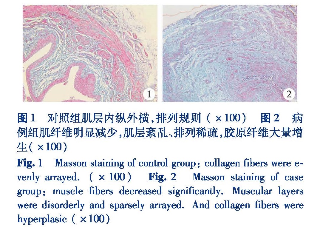
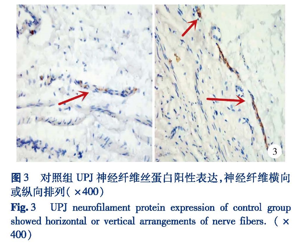
目的 采用病例对照研究的方法来探讨肾盂输尿管连接部Cajal间质细胞(interstitial cell of cajal, ICC)以及代表信息传递的神经组织的改变与先天性肾积水发病机制的关系及其临床意义。方法 收集2013年01月至2014年12月由本院确诊的30例非管腔压迫或新生物所致先天性肾盂输尿管连接部梗阻(Ureteropelvic junction obstruction, UPJO)患儿为病例组; 选择同期11例无肿瘤细胞浸润肾盂输尿管连接部的肾肿瘤患儿作为对照组。所有标本观察组织学改变、神经纤维和ICC标记物的变化。结果 病例组肌层肥厚,排列紊乱、稀疏,其间可见大量增粗胶原纤维。与对照组相比,管腔明显狭窄; 与对照组比较,病例组平滑肌神经纤维丝蛋白大部分标本呈阴性表达,仅少部分标本呈弱阳性表达,且神经纤维异常增粗。病例组神经纤维分布密度为(1.51±0.39),对照组为(3.79±0.48),差异有统计学意义(t=15.36,P<0.01); 病例组UPJ肌层仅见少量ICC呈C-kit阳性表达,甚至部分标本C-kit免疫反应呈阴性表达。病例组ICC标记物分布密度为(1.70±1.24),对照组为(9.09±1.76),差异有统计学意义(t=15.099,P<0.01)。结论 神经纤维与Cajal间质细胞减少在先天性肾盂输尿管连接部梗阻的病因和发病机制中起重要作用,术中应彻底切除肾盂输尿管连接部病变段,以减少术后复发。
Objective To explore the pathogenesis of congenital ureteropelvic junction obstruction(UPJO)in the etiology of congenital hydronephrosis. Methods Thirty specimens of UJPO(without lumen oppression or neoplasm)were collected. And 11 homochronous specimens of UPJ(from cases of renal tumors without an infiltration of tumor cells in UPJ)were selected as controls. The histological changes and distribution density of markers of nerve fibers and ICC were surveyed. Results In case group, there were fibrous hyperplasia, muscular hypertrophy and collagen hyperplasia. Microfilaments proteins were negative in most lesion segments and only few samples showed weak positive expression with abnormal morphology. Few ICCs expressed C-kit in muscular lesion segment of UPJ. Compared with control group, the difference was significant in distribution density of markers(P<0.01). Conclusion A reduction of nerve fibers and ICC may play an important role in the etiology of UPJO. Clinically, ureteropelvic junction stenosis should be totally removed for preventing recurrence.