引言
先天性锁骨假关节(congenital pseudarthrosis of the clavicle,CPC)是一种罕见畸形,1910年由Fitzwilliams首次报道[1-17]。最多见于右锁骨中1/3[4,18-21],约10%累及双侧[14,22],左侧极少受累[7,12,23]。左侧发病者常伴发右位心[9,17,18,23-25]或颈肋[1-6,10,11,14,22,26]。患儿出生时即可发现锁骨干中部有一无痛性包块,随年龄逐渐增大,多数不影响上肢功能[19]。现将我科收治的1例先天性锁骨假关节及文献复习报告如下。
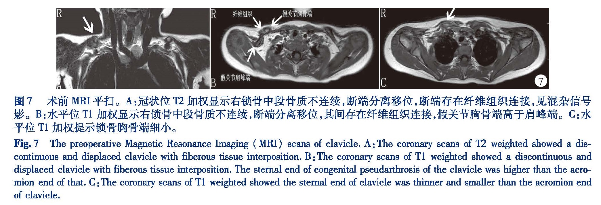
患儿,女,2岁7个月,孕足月剖宫产第一胎,产前检查未见明显异常,无产伤史,无出生窒息缺氧史。生后即发现右锁骨中段包块,质硬,不活动,无压痛,右肩关节和上肢活动正常。生后第4天(图1)﹑第38天(图2)和2岁6个月(图3)的锁骨正位X线片均显示右锁骨干中部分离,断端边缘光滑,肥大呈球状。患儿于2016年3月20日(2岁7个月)来本院就诊。专科检查:右锁骨中段可见一大小约2 cm×2 cm包块(图4),质硬,不活动,无压痛,局部皮肤正常,右肩关节和右上肢活动良好。锁骨X线片显示右锁骨中段骨质不连续,断端分离移位(图5)。CT显示右锁骨中段骨质不连续,断端分离移位,断端间可见软组织密度影(图6)。MRI显示右锁骨中段骨质不连续,断端分离移位,其间见混杂信号影,锁骨胸骨端细小(图7)。
全麻成功后,消毒铺巾。以右锁骨肿物为中心做横行切口,切开皮肤、皮下,切开深筋膜,假关节部位是纤维性组织,无骨性连接,切除假关节,骨缺损约1.0 cm,用直径1.5 mm的克氏针扩髓腔。取左侧髂骨块约1.0 cm×0.8 cm(图8)。克氏针从锁骨肩峰端钻入,将锁骨的外侧部,髂骨骨块和锁骨的内侧部贯穿在一起。在假关节处填置髂骨松质骨骨材。术中C臂透视显示克氏针固定良好(图9)。逐层缝合切口。术后1年随访X线片显示骨性愈合(图10),右肩关节功能正常,无血管神经并发症。
图1 生后第3天X线片显示右侧锁骨于中间断离两部分,断端错位,断端高于锁骨水平;图2生后第38天锁骨正位X线片显示右锁骨中段断裂,断端圆钝﹑移位,且高于锁骨水平;图3患儿2岁6个月时锁骨正位X线片显示右锁骨从中间分离成左右两部分,断端边缘光滑整齐,可见硬化边。
Fig.1 The plain radiogragh at 3 days after brith showed a broken in the middle of the affected clavicle.The fragments were higher than the level of the clavicle;Fig.2 The plain radiogragh at 38 days after brith showed a broken in the middle of the affected clavicle.The fragments were round,displaced,and higher than the clavicle;Fig.3 The plain radiogragh at 30 months old showed the affected clavicle was divided into two fragments.The sclerotic rims were seen at the margins of the fragments。图4 术前外观相:右锁骨中段包块,质硬,无活动,无压痛,局部表皮正常; 图5 术前锁骨正位片显示右锁骨中段骨质中断、明显分离,断端圆钝;图6 术前三维CT表现。A:三维重建显示右锁骨中段骨质不连续,断端分离移位。B:水平位显示右锁骨中段不连续,断端分离移位,断端之间见软组织密度影。
Fig.4 Preoperative appearance.A hard fixed mass can be found in the middle of clavicle.there was no tenderness,and the local skin was normal;Fig.5 The preoperative anteroposterior view radiogragh of the clavicle showed a broken and separated clavicle with round rims;Fig.6 Preoperative appreance of three dimensional Computed Tomography(CT)of clavicle.A:there was a discontinuous and displaced clavicle.B:Horizontal scans showed a discontinuous and displaced clavicle with soft tissue interposiotion.
Fig.4 Preoperative appearance.A hard fixed mass can be found in the middle of clavicle.there was no tenderness,and the local skin was normal;Fig.5 The preoperative anteroposterior view radiogragh of the clavicle showed a broken and separated clavicle with round rims;Fig.6 Preoperative appreance of three dimensional Computed Tomography(CT)of clavicle.A:there was a discontinuous and displaced clavicle.B:Horizontal scans showed a discontinuous and displaced clavicle with soft tissue interposiotion.图7 术前MRI平扫。A:冠状位T2加权显示右锁骨中段骨质不连续,断端分离移位,断端存在纤维组织连接,见混杂信号影。B:水平位T1加权显示右锁骨中段骨质不连续,断端分离移位,其间存在纤维组织连接,假关节胸骨端高于肩峰端。C:水平位T1加权提示锁骨胸骨端细小。
Fig.7 The preoperative Magnetic Resonance Imaging(MRI)scans of clavicle.A:The coronary scans of T2 weighted showed a discontinuous and displaced clavicle with fiberous tissue interposition.B:The coronary scans of T1 weighted showed a discontinuous and displaced clavicle with fiberous tissue interposition.The sternal end of congenital pseudarthrosis of the clavicle was higher than the acromion end of that.C:The coronary scans of T1 weighted showed the sternal end of clavicle was thinner and smaller than the acromion end of clavicle.图8 术中髂骨植骨; 图9术中C臂透视显示克氏针固定良好; 图 10术后12个月X线片提示右锁骨假关节骨性愈合。
Fig.8 Iliac bone grafting in the surgery; Fig.9 The intraoperative image intensifier showed the iliac bone graft and two parts of clavicle were fixed by a kirschner wire; Fig.10 The plain radiogragh of clavicle at 12 months postoperatively showed bony healing of pseudarthrosis.1 讨论
先天性锁骨假关节是一种罕见畸形,发病机制尚未明确[3-5,7,9,10-12,14,16-17,19-20,22-30]。可能是由于颈肋或者垂直方向的肋骨压迫锁骨与第一肋骨之间的锁骨下动脉,锁骨下动脉搏动对锁骨增加压力从而影响锁骨的发育[2,5,7,9,10,12,14-19,25,26,30]。锁骨的发育依靠内侧和外侧的两个分离的骨化中心。在胚胎发育第7周时,锁骨形成第一个骨化骨,两个锁骨骨化中心融合失败也可能是先天性锁骨假关节的另一种原因[1-12,14-16,18,20,22,23,25-27,29,30]。然而,Ogata S[31]研究发现锁骨两个初级骨化中心的结合点位于中、外1/3交界处,而不是锁骨假关节典型的中1/3。Gibson DA[32]认为骨的早期骨化阶段只有一个骨化中心。环境因素在锁骨假关节形成过程中产生了重要作用[3]。先天性锁骨假关节的发生也可能与RUNX2(CBFAI)基因突变相关[6]。
本研究检索了1964—2017年关于先天性锁骨假关节的英文文献(表1)。临床表现主要是锁骨中部无痛性包块[5,18,28]。畸形常伴随患儿逐年加重[7,9,18,26],可出现肩胛骨缩短和下垂。假关节处可由于活动和压力而出现疼痛,但不影响肩关节的正常功能[6,9]。突起处的皮肤可能萎缩变薄,随着上肢的抬高会加重假关节的肿胀[2,5,9]。大体标本表现为锁骨中段不连续,两断端肥厚,有纤维组织分隔。组织学分析显示假关节的近端和远端均有透明软骨帽,软骨帽之间有纤维软骨连接,丰富的胶原纤维组织中存在单个或成对的软骨细胞[11]。
先天性锁骨假关节应与锁骨颅骨发育不良、产道损伤以及多发性神经纤维瘤相鉴别[3,7,10-13,15,19,23,29,30]。先天性锁骨假关节的形成与颈肋关系密切[19]。锁骨颅骨发育不良患儿可见锁骨部分或全部缺失,但无假关节形成,同时伴有颅骨骨化发育不全、赘生牙、身材矮小[1,5,16,23]、脊柱侧凸以及骨盆缺陷[2]。诊断先天性锁骨假关节时应排除外伤史和产伤史,外(产)伤者有局部压痛,X线片可见骨痂形成。
大多数锁骨假关节不影响功能,其治疗方式存在争议。若无局部美观的要求,同时患侧肩和上肢无临床症状者,可选择保守治疗[2,12,19,28,33]。手术治疗先天性锁骨假关节可以完全恢复肩关节活动的生理功能,预防胸廓出口综合征,改善美观[2,7-9,12,19,23,29]。
先天性锁骨假关节最常见的并发症为胸廓出口综合征,是由于锁骨下动脉(动脉性胸廓出口综合征)、锁骨下静脉和臂丛神经(神经性胸廓出口综合征)在胸廓上口受压迫而导致上肢和手掌发凉、疲劳、局部贫血、坏疽、血栓栓塞、锁骨下动静脉瘤、水肿和感觉异常等[3]。手术方式主要是锁骨假关节切除术结合骨移植术和内固定术[1-4,8,10,12,17,19-20,24,26,27,33]。手术年龄一般为在3~14岁,以5~7岁为最佳,可以避免肩部缩短和生长不对称[7]。骨移植物可以是自体骨、同种异体骨和自体干细胞[6],以自体髂骨为最佳[6,34]。取自体髂骨的主要并发症是疼痛[4]。内固定物的选择可以是克氏针、施氏针、螺纹针、加压钢板或者外固定架[4,8,9,19]。加压钢板能够更快促进骨性愈合[8],优于螺纹针带来的术后高感染发生率和假关节不愈合发生率。术后并发症包括假关节不愈合、延迟愈合、局部感染、脓毒症、肩部瘢痕和臂丛经损伤等[2]。
-
1 Sung TH,Man EM,Chan AT,et al.Congenital pseudarthrosis of the clavicle:a rare and challenging diagnosis [J].Hong Kong Med J,2013,19(3):265 — 267.DOI:10.12809/hkmj133648.
-
2 Galanopoulos I,Ashwood N,Garlapati AK,et al.Congenital pseudarthrosis of clavicle:illustrated operative technique and histological findings [J].BMJ Case Rep,2012,DOI:10.1136/bcr — 2012 — 006908.
-
3 Watson HI,Hopper GP,Kovacs P.Congenital pseudarthrosis of the clavicle causing thoracic outlet syndrome [J].BMJ Case Rep,2013,2013.pii:bcr2013010437.DOI:10.1136/bcr — 2013 — 010437.
-
4 Elliot RR,Richards RH.Failed operative treatment in two cases of pseudarthrosis of the clavicle using internal fixation and bovine cancellous xenograft(Tutobone)[J].J Pediatr Orthop B,2011,20(5):349 — 353.DOI:10.1097/BPB.0b013e328346c010.
-
5 Currarino G,Herring JA.Congenital pseudarthrosis of the clavicle [J].Pediatr Radiol,2009,39(12):1343 — 1349.DOI:10.1007/s00247 — 009 — 1396 — 1.
-
6 Di Gennaro GL,Cravino M,Martinelli A,et al.Congenital pseudarthrosis of the clavicle:a report on 27 cases [J].J Shoulder Elbow Surg,2017,26(3):e65 — e70.DOI:10.1016/j.jse.2016.09.020.
-
7 Studer K,Baker MP,Krieg AH.Operative treatment of congenital pseudarthrosis of the clavicle:a single-centre experience [J].J Pediatr Orthop B,2017,26(3):245 — 249.DOI:10.1097/BPB.0000000000000400.
-
8 Chandran P,George H,James LA.Congenital clavicular pseudarthrosis:comparison of two treatment methods [J].J Child Orthop,2011,5(1):1 — 4.DOI:10.1007/s11832 — 010 — 0313 — 3.
-
9 Figueiredo MJ,Dos Reis Braga S,Akkari M,et al.Congenital pseudarthrosis of the clavicle [J].Rev Bras Ortop,2015,47(1):21 — 26.DOI:10.1016/S2255 — 4971(15)30341 — 4.
-
10 Persiani P,Molayem I,Villani C,et al. Surgical treatment of congenital pseudarthrosis of the clavicle:a report on 17 cases [J].Acta Orthop Belg,2008,74(2):161 — 166.
-
11 Gomez-Brouchet A,Sales de Gauzy J,Accadbled F,et al.Congenital pseudarthrosis of the clavicle:a histopathological study in five patients [J].J Pediatr Orthop B,2004,13(6):399 — 401.
-
12 Ettl V,Wild A,Krauspe R,et al.Surgical treatment of congenital pseudarthrosis of the clavicle:a report of three cases and review of the literature [J].Eur J Pediatr Surg,2005,15(1):56 — 60.DOI:10.1055/s — 2004 — 817944.
-
13 Sales de Gauzy J,Baunin C,Puget C,et al.Congenital pseudarthrosis of the clavicle and thoracic outlet syndrome in adolescence [J].J Pediatr Orthop B,1999,8(4):299 — 301.
-
14 Sakkers RJ,Tjin a Ton E,Bos CF.Left-sided congenital pseudarthrosis of the clavicula [J].J Pediatr Orthop B,1999,8(1):45 — 47.
-
15 Schnall SB,King JD,Marrero G.Congenital pseudarthrosis of the clavicle:a review of the literature and surgical results of six cases [J].J Pediatr Orthop,1988,8(3):316 — 321.
-
16 Quinlan WR,Brady PG,Regan BF.Congenital pseudarthrosis of the clavicle [J].Acta Orthop Scand,1980,51(3):489 — 492.
-
17 Manashil G,Laufer S.Congenital pseudarthrosis of the clavicle:report of three cases [J].AJR Am J Roentgenol,1979,132(4):678 — 679.DOI:10.2214/ajr.132.4.678.
-
18 温树正,郭文通,李文琪,等.先天性锁骨假关节 1 例报告[J].内蒙古医学院学报,1997,19(4):25.DOI:10.16343/j.cnki.issn.2095 — 512x.1997.04.010.
-
19 Glotzbecker MP,Shin EK,Chen NC,et al.Salvage reconstruction of congenital pseudarthrosis of the clavicle with vascularized fibular graft after failed operative treatment:a case report [J].J Pediatr Orthop,2009,29(4):411 — 415.DOI:10.1097/BPO.0b013e3181a5ebff.
-
20 Brévaut-Malaty V,Guillaume JM.Neonatal diagnosis of congenital pseudarthrosis of the clavicle [J].Pediatr Radiol,2009,39(12):1376.DOI:10.1007/s00247 — 009 — 1424 — 1.
-
21 Cataldo F.A 7-month-old child with a clavicular swelling since birth.Diagnosis:congenital pseudarthrosis of the clavicle [J].Eur J Pediatr,1999,158(12):1001 — 1002.
-
22 Grogan DP,Love SM,Guidera KJ,et al.Operative treatment of congenital pseudarthrosis of the clavicle [J].J Pediatr Orthop,1991,11(2):176 — 180.
-
23 O'Leary E,Elsayed S,Mukherjee A,et al.Familial pseudarthrosis of the clavicle:does it need treatment ? [J].Acta Orthop Belg,2008,74(4):437 — 440.
-
24 Sforza G,Levy O.Posttraumatic locked dislocation of congenital pseudarthrosis of the clavicle[J].J Shoulder Elbow Surg,2005,14(3):336 — 337.DOI:10.1016/j.jse.2004.06.017.
-
25 Russo MT,Maffulli N.Bilateral congenital pseudarthrosis of the clavicle [J].Arch Orthop Trauma Surg,1990,109(3):177 — 178.
-
26 Morin LR,Fossey FP,Besselièvre A,et al.Congenital pseu-darthrosis of the clavicle[J].Acta Obstet Gynecol Scand,1993,72(2):120 — 121.
-
27 Nieto Gil A,Gómez Navalón A,Zorrilla Ribot P.Bilateral congenital seudarthrosis of the clavicle [J].A clinical case,Rev Esp Cir Ortop Traumatol,2016,60(6):397 — 399.DOI:10.1016/j.recot.2015.02.004.
-
28 Karakurt L,Yilmaz E,Belhan O,et al.Pycnodysostosis associated with bilateral congenital pseudarthrosis of the clavicle [J].Arch Ortho Trauma Surg,2003,123(2 — 3):125 — 127.DOI:10.1007/s00402 — 003 — 0484 — 1.
-
29 Lorente Molto FJ,Bonete Lluch DJ,Garrido IM.Congenital pseudarthrosis of the clavicle:a proposal for early surgical treatment [J].J Pediatr Orthop,2001,21(5):689 — 693.
-
30 Hirata S,Miya H,Mizuno K.Congenital pseudarthrosis of the clavicle.Histologic examination for the etiology of the disease [J].Clin Orthop Relat Res,1995,(315):242 — 245.
-
31 Ogata S,Uhthoff HK.The early development and ossification of the human clavicle-an embryologic study [J].Acta Orthop Scand,1990,61(4):330 — 334.
-
32 Gibson DA,Carroll N.Congenital pseudoarthrosis of the clavicle [J].J Bone Joint Surg,1970,52(4):644 — 652.
-
33 Shalom A,Khermosh O,Wientroub S.The natural history of congenital pseudarthrosis of the clavicle [J].J Bone Joint Surg Br,1994,76(5):846 — 847.
-
34 Heidt C,Ziebarth K,Erni D,et al.Four years follow-up after clavicle reconstruction in a child:a case report [J].Plast Reconstr Aesthet Surg,2014,67(12):1735 — 1739.DOI:10.1016/j.bjps.2014.08.050.
-
35 Morrison MC.Congenital Pseudarthrosis of Clavicle [J].Proc R Soc Med,1964,57(2):94.


