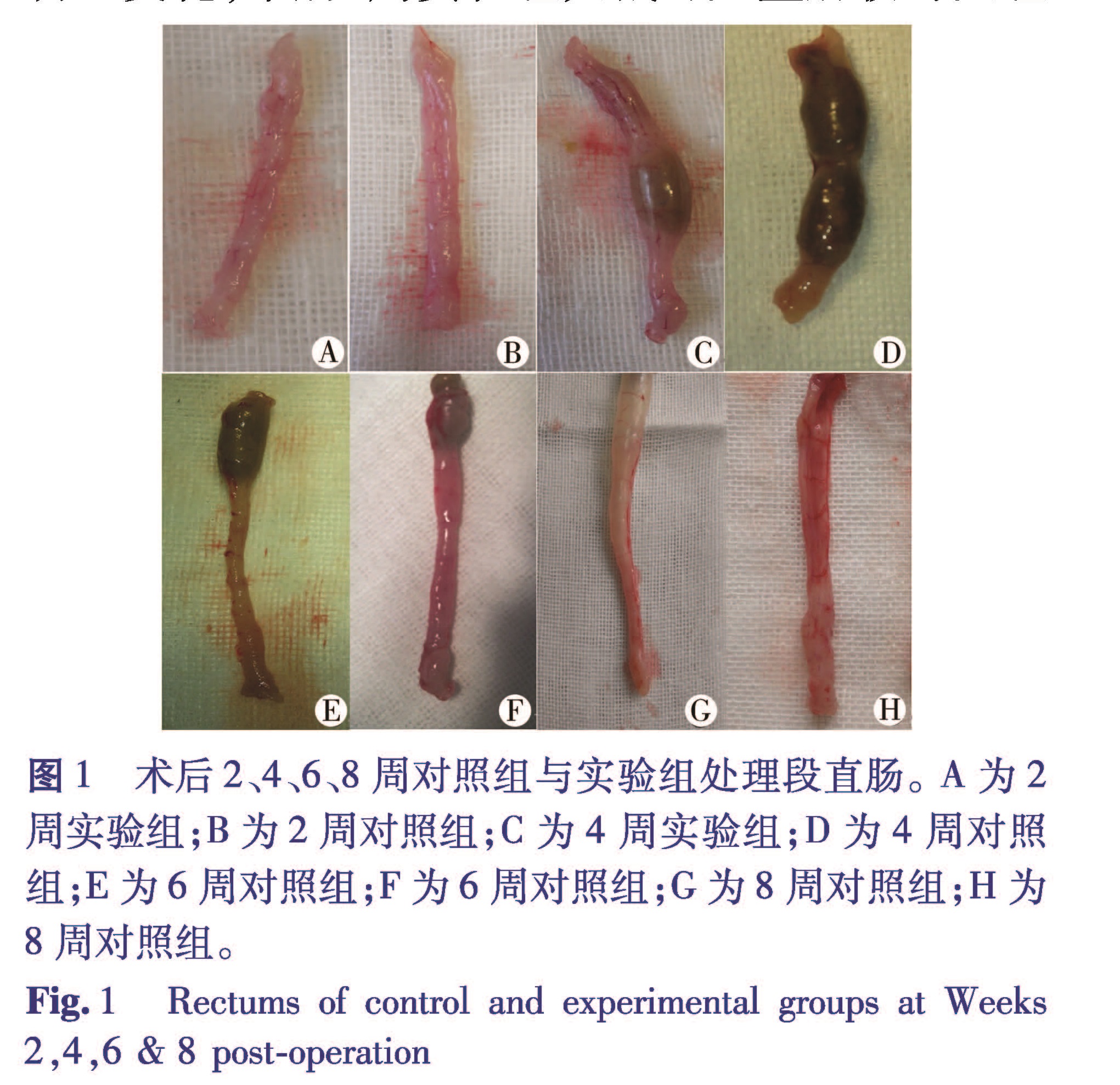
近年来,国内外学者已从胚胎和成体组织成功提取出肠神经嵴干细胞(ENCSCs),并在体外培育成神经球(NLBs),且该NBLs可分化为神经元和神经胶质细胞,这为移植替代治疗HD提供了可靠的供体[1-5]。但早期的HD移植模型多为体外培育的肠管组织和基因修饰的动物模型[6],体外培养组织移植存在局限性,而基因修饰模型虽然能造成肠道神经节的缺如,但实验动物大多因基因的缺陷出生不久后死亡,二者均较难观察成体肠管内移植后的情况[7]。1978年Sato A[8]第一次报道使用BAC破坏肠道神经元,成功建立HD大鼠模型。此后,国内外学者也成功建立了空肠、回肠、结肠无神经节细胞症的动物模型[9,10]。但以上几种模型均需行开腹关腹手术,如需行移植实验,还需要二次手术,模型死亡率高,故本实验通过直肠灌注BAC法,避免了开腹关腹的风险,明显提高了术后存活率,且采用乳鼠作为实验对象,更加符合小儿HD的特点,这便成为了一种更加合适的移植模型。
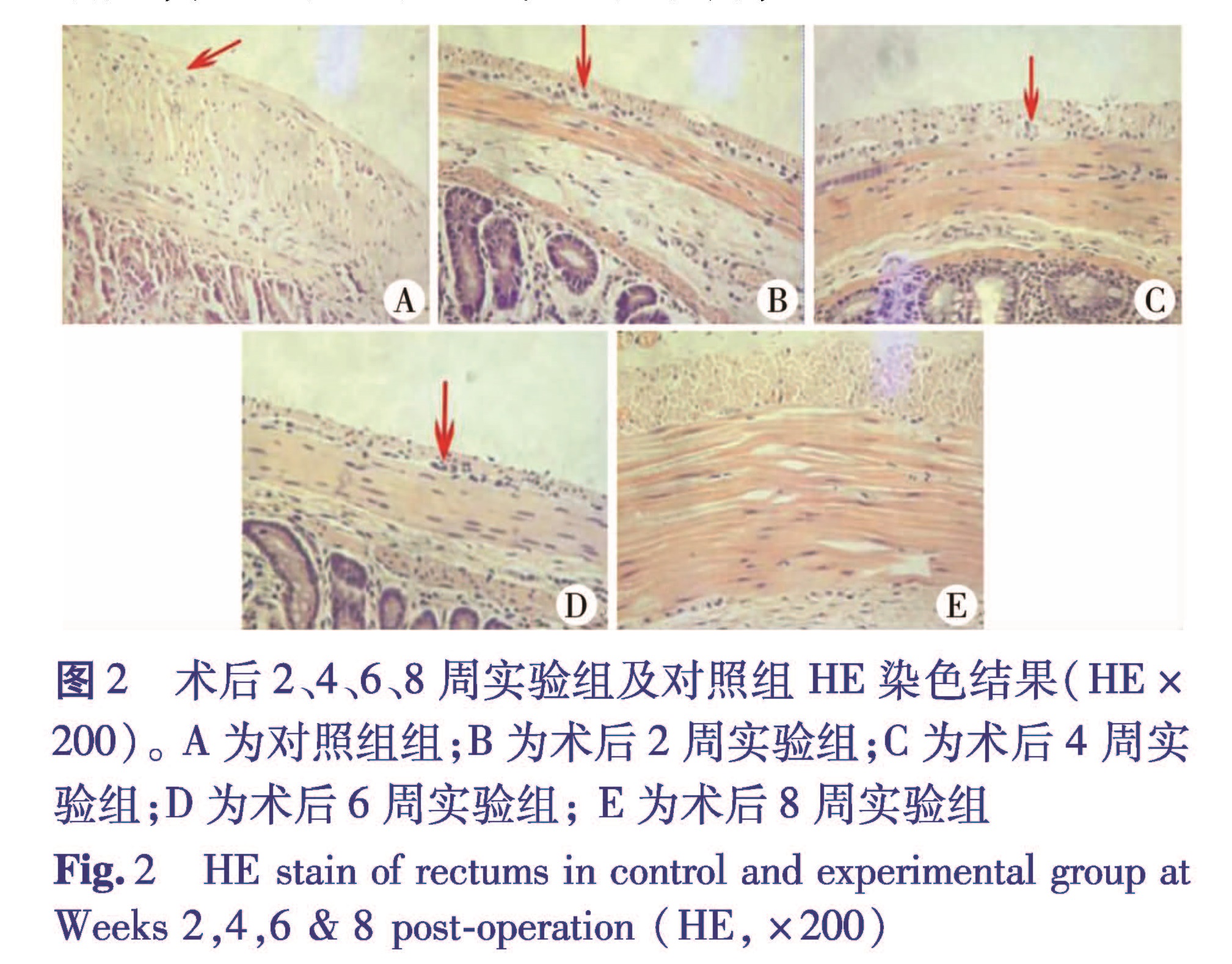
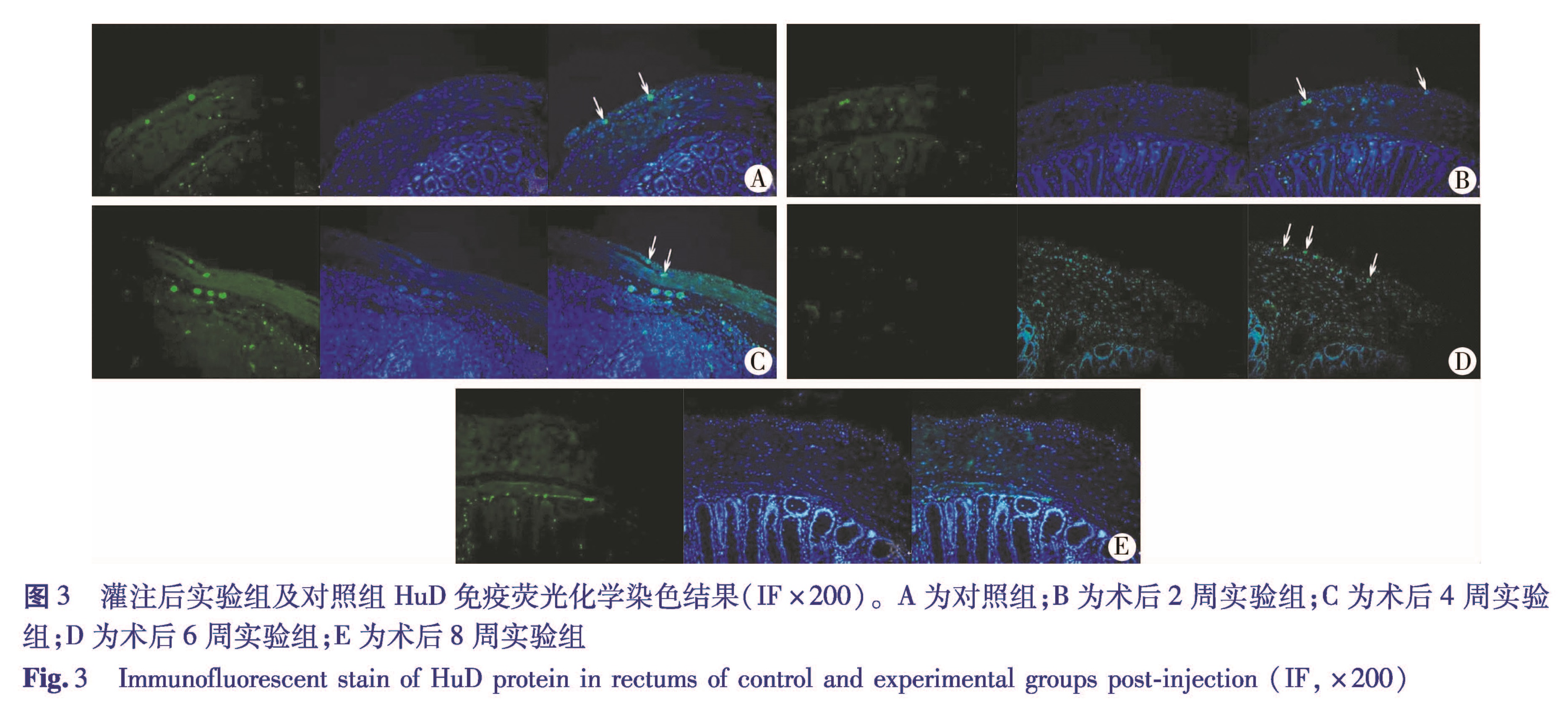
HuD蛋白是一种RNA结合蛋白,可以清晰的显示出肠道肌间神经丛的神经节细胞,它与其他标记物不同,HuD多在核内表达,胞质基本无表达,神经纤维基本不着色,因此在行免疫荧光染色时,可以有效地分辨出神经元的数量、大小,排除了神经纤维的干扰[11]。本研究显示6周时实验组HuD的肌间神经丛变小、形态各异,至8周时已无法看到HuD表达。可以看出,直肠灌注BAC可以导致大鼠直肠神经节细胞完全缺如,达到与临床上HD相似的效果。
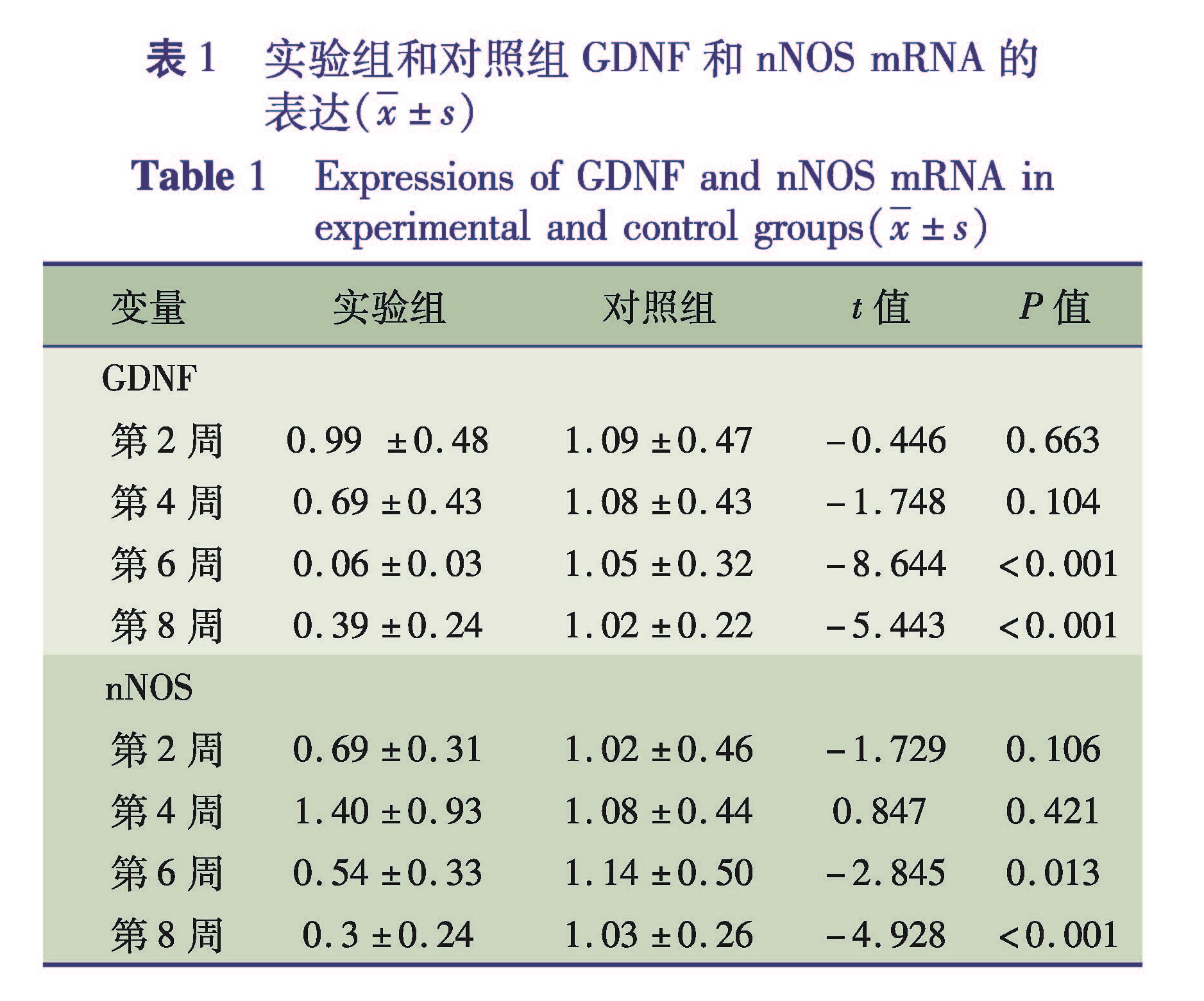
GDNF是一种分泌蛋白,属于转化生长因子β超家族,通过与GDNF家族受体α1结合形成复合物,可以结合并激活跨膜受体酪氨酸激酶(RET),促进胚胎期肠ENCSCs的迁移、增殖、分化、存活而形成正常的ENS,胚胎期GDNF的缺乏将导致ENS发育缺陷,引起HD的发病[12,13]。目前国内外学者在对HD患儿病变肠管GDNF表达的研究中发现患儿病变肠管的表达量较正常组均降低,这与我们的动物实验结果一致[14,15]。值得注意的是,GDNF在8周实验组较6周实验组明显增高,原因不明,查阅相关文献认为是GDNF在成体神经元的轴突生长、细胞存活及神经损伤再修复上起到重要的作用,BAC处理后的肠管在一段时间后,肠神经系统存在自我修复情况,但这种自我修复的机制目前尚不清楚[16-20]。
nNOS是肠道合成释放抑制性神经递质NO的关键酶,当缺乏这种关键酶时,NO的合成释放将会减少,可导致在平滑肌水平缺乏一个抑制性松弛信号,使NO缺乏段肠管紧张度增加。肠道内NO由肌间神经节通过激活的nNOS合成后释放,肠管肌间神经节可以检测到nNOS的表达,先天性巨结肠患者病变段的nNOS mRNA 表达是减少的甚至不表达,我们的结果也显示了nNOS mRNA在实验组的表达降低,这也进一步表明BAC处理后的肠管能够建立与临床HD类似的无神经节细胞动物模型 [21,22]。但是我们所制作的这种无神经节细胞大鼠模型,尚不能完全代替临床上的HD,相对而言,基因敲除动物模型可以达到相对完善的HD模型,但其制作难度及成本高、实验条件苛刻,不能普及,而我们所建立的HD模型存活率高,存活时间长,饲养方便,更适合研究无神经节细胞性巨结肠病的病理过程、发病机制和治疗措施。