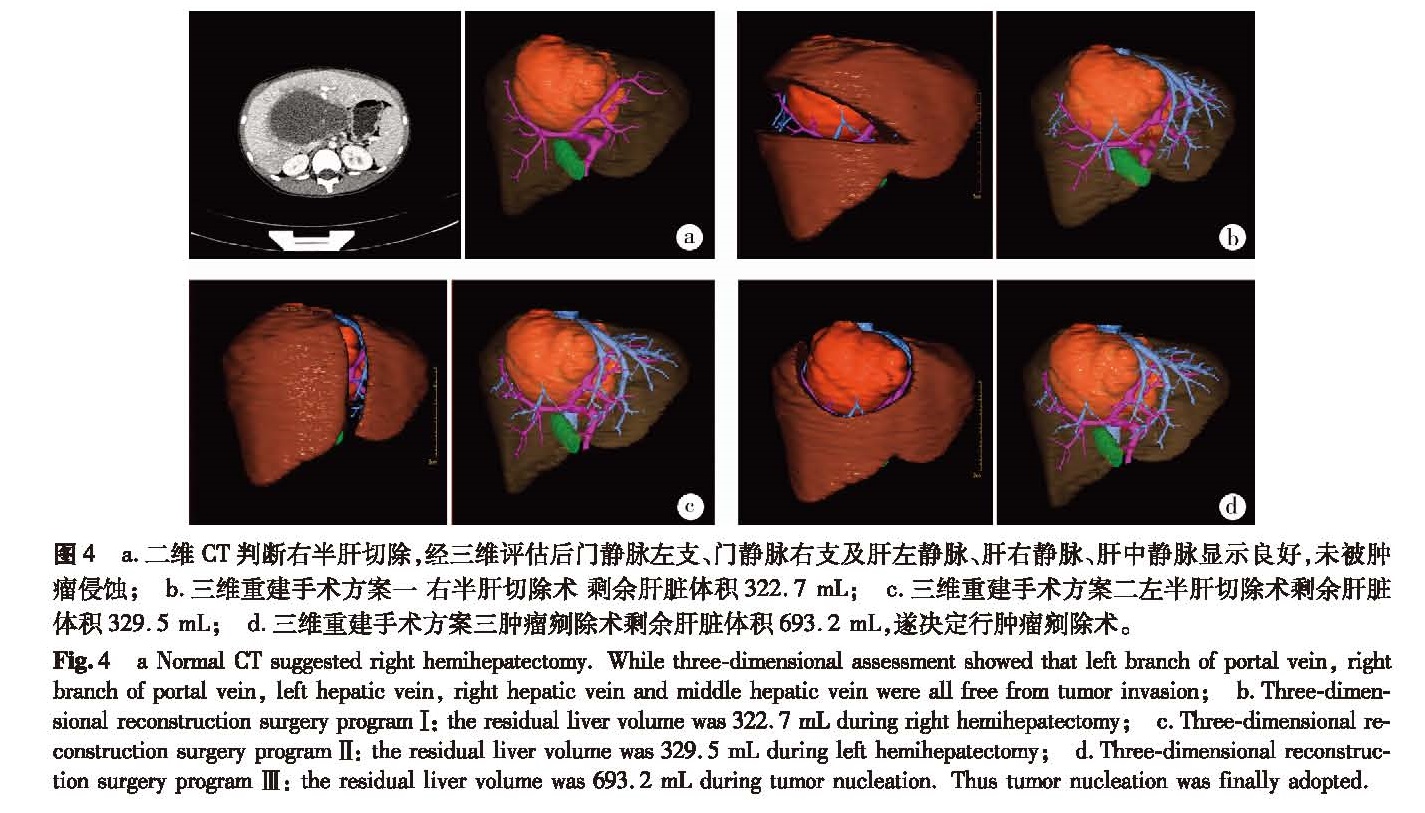
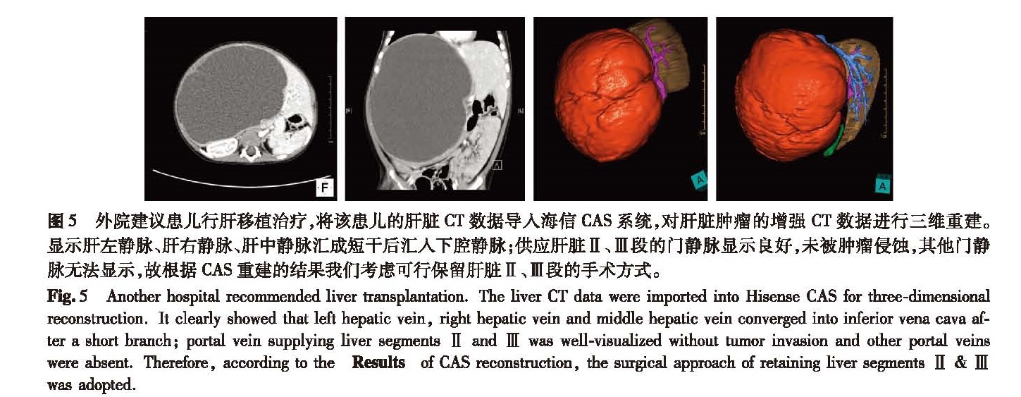
目的 对儿童肝脏肿瘤与门静脉重要分支的位置关系、压迫、侵袭情况进行医学数字影像分析,探讨海信CAS系统在儿童肝脏肿瘤手术中的临床价值。 方法 收集2015年1月至2016年12月间由青岛大学附属医院及浙江大学医学院附属儿童医院收治的56例肝脏肿瘤患儿的CT数据。其中男童30例,女童26例,年龄1个月至12岁,平均年龄5.5岁; 肝母细胞瘤42例,肝细胞癌5例,婴儿型血管内皮细胞瘤5例,间叶性错构瘤1例,未分化肉瘤1例,畸胎瘤肝转移2例; 运用海信CAS系统对其增强CT数据进行三维重建,对肝脏肿瘤及门静脉分支的位置关系、压迫、侵蚀情况进行分析,虚拟手术切除肿瘤,测量残余肝脏体积并计算残余肝脏体积百分比。 结果 56例患儿中,肿瘤位于肝左叶7例,其中肿瘤侵犯门静脉左支主干(门静脉2级分支)6例,肿瘤侵犯门静脉左支3级分支1例; 肿瘤位于肝右叶40例,其中肿瘤侵犯门静脉右支主干(门静脉2级分支)33例,肿瘤侵犯门静脉右支3、4级分支7例; 肿瘤位于肝中叶8例,其中肿瘤侵蚀门静脉左支、右支主干(门静脉2级分支)5例,肿瘤未侵蚀门静脉左支、右支主干(门静脉2级分支),仅对门静脉左支、右支主干(门静脉2级分支)形成压迫3例; 全肝病变1例。 结论 利用海信CAS系统进行三维重建所得的数字模型能够清晰、准确地显示小儿肝脏肿瘤与门静脉分支的关系,对儿童肝脏手术有重要的指导意义。
Objective To analyze the relationship between liver tumors and portal veins and explore the clinical value of Hisense computer-assisted surgery system(CAS)during pediatric liver surgery. Methods The clinical data of 56 cases with liver tumors from January 2015 to December 2016 was analyzed. There were 30 boys and 16 girls with an age range of 1 month to 12 years. There were hepatoblastoma(n=42), hepatocellular carcinoma(n=5), hemangioendothelioma(n=5), mesenchymalhamartoma(n=1), undifferentiated sarcoma(n=1)and hepatic metastases of teratoma(n=2). Hisense CAS was used for three-dimensional reconstructing based upon thin-layer computed tomography(CT). Contrast operation planning was based on common CT imaging along with three-dimensional reconstructing results. Residual liver volume was measured after virtual tumor resection. Results In left lobe of liver(n=7), tumors invaded left portal vein trunk(secondary branches of portal vein)(n=6)and the third branches of portal vein(n=1); in right lobe of liver(n=40), tumors involved the right portal vein trunk(secondary branches of portal vein)(n=33)and the third or forth branches of portal vein(n=7); in medial lobe of liver(n=8), tumors infiltrated right or left portal vein trunk(secondary branches of portal vein)(n=5)and right or left portal vein trunk(secondary branches of portal vein)(n=3); diffuse liver tumor(n=1). Six cases had no chance of one-stage surgery. Conclusion Hisense CAS can delineate distinctly vascular system and adjacent relationship of tumor so as to improve the accuracy and safety during pediatric liver surgery.