小儿四肢骨缺损传统治疗方法多采用自体骨或异体骨充填移植。儿童时期可供移植用的骨量较少,当骨缺损病变范围较大时,自体骨无法满足移植的需要,且术后可造成取骨部位慢性疼痛、切口感染等; 异体骨诱导性差,有传播疾病的风险[4,7]。近年来,骨组织工程技术有望成为修复大段骨缺损的有效途径之一,建立幼龄动物长骨大段骨缺损动物模型对于研究儿童骨缺损修复的实验研究具有重要的意义。
骨缺损实验中,常用动物有羊、犬、猪,鼠、兔等。鼠类虽然实验成本较低,但其骨结构原始,骨损伤后修复机制与人类不同[3]。兔类具有与人体相似的骨组织结构,建模后能较好地模拟人骨缺损后的生物力学改变及骨修复机制。新西兰兔桡骨结构与人类相似,上下尺桡关节连接紧密,骨干间由骨间膜紧密结合,活动度小。且桡骨不是主要负重骨,制造骨缺损后,临近尺骨可支撑固定,不需额外植入内外固定物,能够避免术后负重及固定物对骨愈合的影响,对动物日常活动影响较小[4,8]。
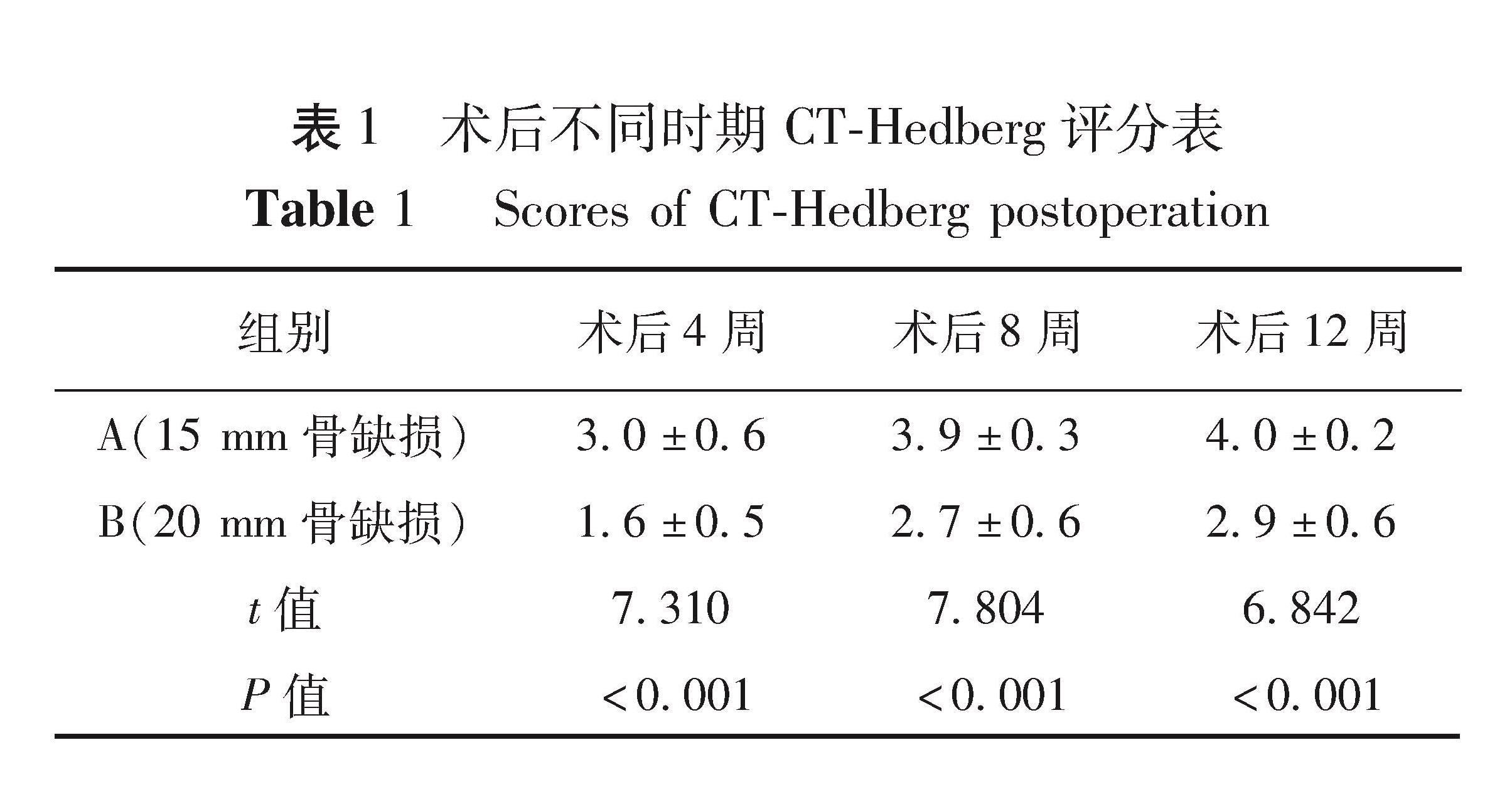
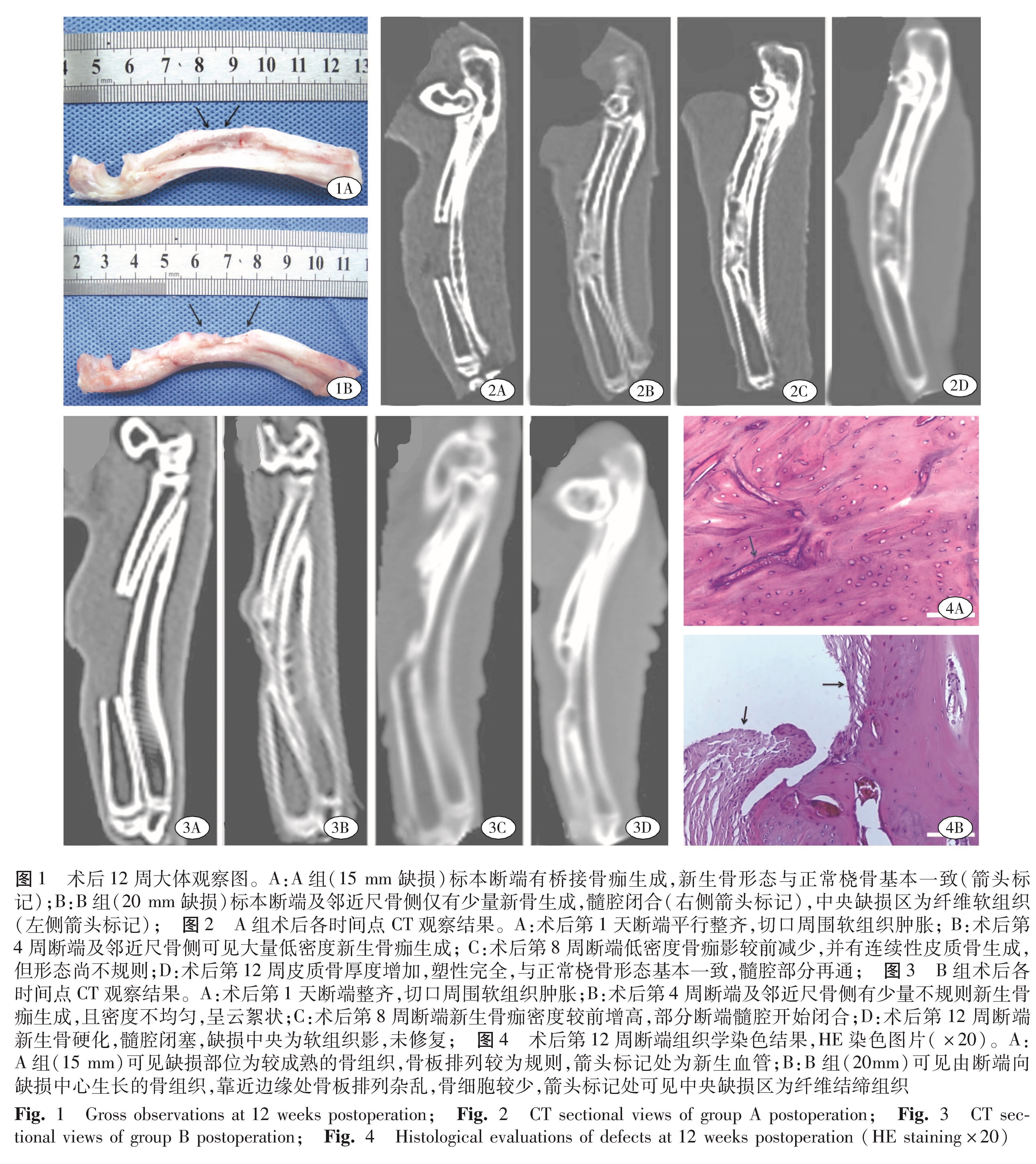
合适的大段骨缺损尺寸,即临界性骨缺损(critical size defect,CSD),是动物骨缺损模型的重要参数,它指在一定周期内未经治疗、无法自愈的最短骨缺损长度[9]。有学者认为动物长骨CSD长度应为其周径的1.5~2.5倍以上或为长骨长度的10%以上[10]。既往研究中存在多种长骨CSD大小,从10~20 mm不等[8,11]。参考李东亚等[11]在6月龄新西兰兔桡骨制备骨缺损的方法,并结合本研究中对兔桡骨影像学相关测量数据,显示桡骨中段周径约为9 mm,本实验选择了15 mm及20 mm两种骨缺损尺寸,并未设置更大长度缺损组。本实验CT结果显示:术后第8周15 mm组绝大部分断端有桥接骨痂形成,而20 mm组标本仍存在缺损空腔; 术后第12周,15 mm组断端基本塑形完全,与正常桡骨形态大致相同,20 mm组断端髓腔闭合,中央缺损区未见连续性骨痂生长。且术后各时间点,15 mm组CT-Hedberg评分均高于20 mm组,差异有统计学意义。Meimandi等[8]在实验中选取10~12月龄雄性新西兰兔制作10 mm桡骨干骨缺损模型,术后60 d后组织学结果显示,断端仅有纤维性连接,尚未形成桥接骨痂,且三点弯曲试验显示其生物力学稳定性较正常骨组织差。李东亚等[11]对桡骨骨缺损大小进行了对照研究,他们选取18只雄性6月龄新西兰兔随机分为3组分别制作10 mm、15 mm、20 mm桡骨干骨缺损,术后12周大体标本显示10 mm组断端愈合完全,而15 mm、20 mm组均未愈合,缺损空腔由纤维组织填充,Hedberg评分显示15 mm组评分显著小于10 mm组,而与20 mm组相比,二者间差异无统计学意义,基于该实验结果,他们认为6月龄雄性新西兰兔桡骨CSD大小为15 mm较合理。Bodde EW等[6]认为实验动物月龄是影响骨再生的一个重要因素。3月龄新西兰兔处于幼龄期,约相当于人类儿童5~7岁[12]。Rivas研究发现新西兰兔长骨在19~32周时停止生长[12]。本实验骨缺损愈合时间及CSD大小与Meimand Parizi A [8]及李东亚等[11]国内外实验结果存在差异,可能是因为本研究选择了骨生长旺盛的幼龄期新西兰兔,而后两者选择了较为成熟的6月龄成年兔。王永刚等[13]选择4~5月龄新西兰兔制作股骨干骨缺损,直到术后12周断端才形成骨痂桥接; Song等[14]选择构建6周龄新西兰兔股骨干骨缺损模型,术后5~6周X线显示断端有大量骨痂生成,术后7~9周即形成骨痂桥接,平均骨愈合时间约为7.3周,短于本研究中骨愈合时间。
沈凯等[15]在5~6月龄新西兰兔桡骨骨缺损模型建模中,保留了骨膜,并设计10 mm、15 mm、20 mm、25 mm及30 mm组进行对照研究,结果发现,20 mm缺损组出现骨愈合。本研究结果与沈凯等[15]的不同,本研究中20 mm组断端并未愈合,可能与术中对缺损两端骨膜进行一定程度的剥离有关。有实验研究发现,桡骨断端骨膜可促进断端骨愈合,应彻底清理断端残余骨膜,以排除其对断端骨愈合的影响[6]。Zhang YD等建议术中截断桡骨后,两侧断端骨膜应向干骺端剥离约5 mm长,并彻底冲洗断端残余骨膜组织[5]。
综上所述,本研究所选择尺寸易于控制,手术操作简单,易于推广。选取3月龄幼兔构建桡骨干骨缺损模型时,骨缺损长度应选择20 mm较为合适。不足之处在于: 选择3月龄幼兔作为实验对象,相关实验结论类比应用于人类患儿其模型代表性存在争议; 实验分组不够详细,考虑到幼兔的实验饲养困难性问题,没有设置1月龄及2月龄组进行对比以探讨不同月龄对骨缺损愈合的影响; 为便于手术操作及误差控制,未细化设置12 mm、14 mm、18 mm组等组别。