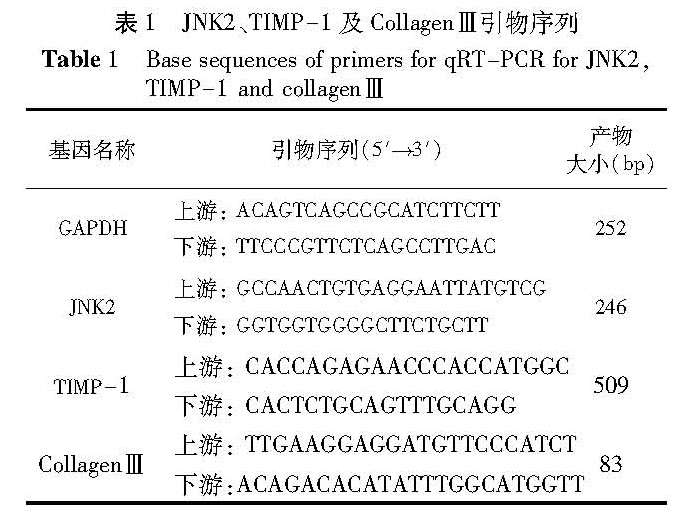
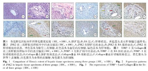
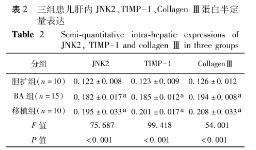
目的 研究c — Jun氨基末端激酶2(c — Jun N-terminal kinase, JNK2)、金属蛋白酶组织抑制剂 — 1(tissue inhibitor of metalloproteinase 1, TIMP — 1)及CollagenⅢ 在胆道闭锁(BA)肝组织中的表达情况及在肝纤维化进程中的作用。 方法 选取胆管扩张症肝活检病例10 例,为胆扩组; BA肝活检病例15 例,为BA组; BA晚期因肝功能衰竭而行肝移植患者自体肝活检10例,为肝移植组。采用HE染色观察并评价肝标本纤维化程度; 免疫组化染色检测JNK2、TIMP — 1及CollagenⅢ在肝组织中的表达; 实时荧光定量聚合酶链式反应(qRT — PCR)检测肝组织中JNK2、TIMP — 1及CollagenⅢ基因表达情况。 结果 ①HE染色: 胆扩组偶可见少许纤维细胞增生; BA组汇管区增宽,纤维组织增生、桥接纤维化现象普遍,可见假小叶形成; 肝移植组汇管区明显增宽,纤维组织增生较重,广泛桥接纤维组织形成,假小叶显著。②免疫组化:胆扩组JNK2、TIMP — 1及CollagenⅢ表达为弱阳性,BA组及肝移植组JNK2、TIMP — 1及CollagenⅢ蛋白均在肝细胞、汇管区胆管上皮细胞及血管内皮细胞胞质中阳性表达。③半定量分析:三组JNK2(0.122±0.008、0.182±0.017 和0.198±0.033),TIMP — 1(0.123±0.009、0.185±0.012和0.201±0.017)和CollagenⅢ(0.126±0.012、0.194±0.008和0.208±0.033)表达比较,差异有显著统计学意义(P<0.001); 三组中,BA组及肝移植组JNK2、TIMP-1及CollagenⅢ表达明显高于胆扩组(P<0.05); 肝移植组JNK2、TIMP — 1及CollagenⅢ蛋白含量与BA组比较,差异无统计学意义(P值均>0.05)。④qRT — PCR:三组JNK2、TIMP — 1和CollagenⅢmRNA表达水平比较,差异有统计学意义(0.221(0.17)vs 1.395(1.22)vs 1.095(1.21),H=17.686,P=0.003; 0.439(0.31)vs 1.404(0.85)vs 1.571(0.66),H=20.648,P=0.000; 0.917(0.09)vs 1.802(1.35)vs 1.957(1.30),H=15.555,P=0.007),BA组及肝移植组肝内JNK2、TIMP — 1及CollagenⅢmRNA表达含量比胆扩组高(P值均小于0.017)。 结论 JNK2、TIMP — 1及CollagenⅢ在BA患儿随着肝纤维化加重而表达升高,表明其可能参与并促进BA肝纤维化进程。
Objective To explore the expressions of c-Jun N-terminal kinase(JNK2), tissue inhibitor of metalloproteinase — 1(TIMP — 1)and collagen Ⅲ in liver tissues and elucidate their functions in the process of liver fibrosis of biliary atresia(BA). Methods Liver biopsy specimens were collected from congenital biliary dilatation(CBD group, n=10), BA liver biopsy(BA group, n=15), BA children undergoing liver transplantation due to liver failure(liver transplantation group, n=10). Hematoxylin and eosin(HE)staining was used for evaluating the degree of liver fibrosis. And the expressions of JNK2, TIMP — 1 and collagen Ⅲ in liver tissue were detected by immunohistochemical staining. Quantitative real-time polymerase chain reaction(qRT — PCR)was used to test the gene expressions of JNK2, TIMP-1 and collagen Ⅲ. Results ①HE staining: Fiber cell hyperplasia in CBD group; Portal area expansion, fibrous tissue proliferation, bridging fibrosis and little few pseudo lobules in Kasai group; Portal area became widened obviously, fibrous tissue proliferation was heavier, bridging fibrosis generally formed, pseudo-lobular was remarkable in liver transplantation group. ②Immunohistochemistry: The expressions of JNK2, TIMP-1 and collagenⅢ were weakly positive in CBD group. The positive expression of JNK2, TIMP — 1 and collagen Ⅲ protein in hepatocytic cytoplasm, portal area of bile duct epithelial cells and vascular endothelial cells in BA and transplantation groups. Semi-quantitative analysis: The expression levels of JNK2, TIMP — 1 and collagen Ⅲ protein had significant differences among three groups(0.122±0.008 vs 0.182±0.017 vs 0.198±0.033, F=75.687, P=0.000; 0.123±0.009 vs 0.185±0.012 vs 0.201±0.017, F=99.418, P=0.000; 0.126±0.012 vs 0.194±0.008 vs 0.208±0.033, F=54.001, P=0.000); BA and liver transplantation groups were significantly higher than that in CBD group(P<0.05). No significant differences existed in protein level between BA and transplantation groups(P>0.05); qRT — PCR: The mRNA expression levels of JNK2, TIMP — 1 and collagenⅢ had significant differences among three groups(0.221(0.17)vs 1.395(1.22)vs 1.095(1.21), H=17.686, p=0.003; 0.439(0.31)vs 1.404(0.85)vs 1.571(0.66), H=20.648, P=0.000; 0.917(0.09)vs 1.802(1.35)vs 1.957(1.30), H=15.555, P=0.007); The mRNA expression of JNK2, TIMP-1 and collagen Ⅲ were higher in BA group and liver transplantation group than those of CBD group(P<0.017). Conclusion s The expressions of JNK2, TIMP — 1 and Collagen Ⅲ increased in liver of BA during fibrosis. It hints that the expressions of JNK2, TIMP — 1 and collagen Ⅲ may promote the process of liver fibrosis in BA.
![表3 三组患儿中 JNK2、TIMP — 1, Collagen Ⅲ mRNA 相对含量比较[M(IQR)]<br/>Table 3 Comparison of expression levels of JNK2 etc. mRNA between three groups M[(IQR)]](2017年02期/pic07.jpg)