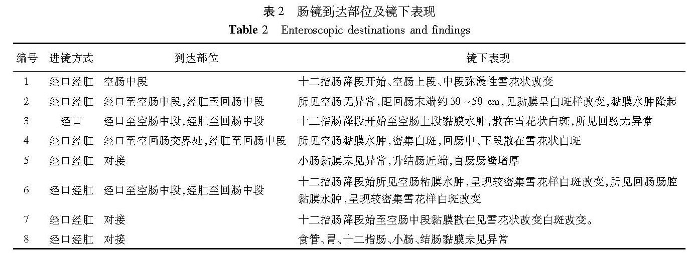
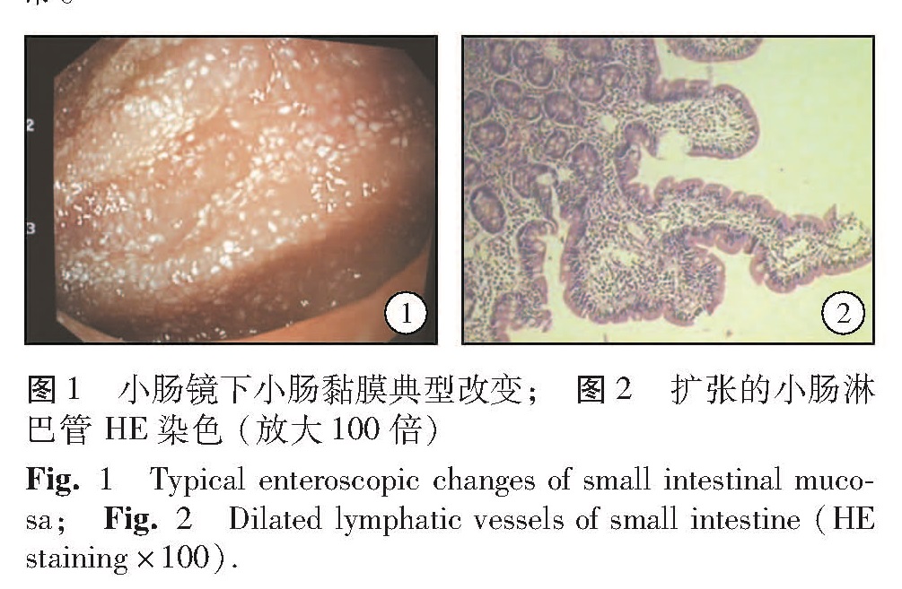
目的 探讨单气囊小肠镜检查+小肠黏膜活检对儿童原发性小肠淋巴管扩张(primary intestinal lymphangiectasia,IL)的诊断价值。 方法 采用奥林巴斯SIF — Q260型SBE对2009 年5 月至2015年4 月我们收治的肠淋巴管扩张症患儿,进行单气囊小肠镜检查和小肠黏膜活检。 结果 8例患者共接受了15次单气囊小肠镜检查,经口腔8次,经肛门7次,平均检查时间120 min(95~180 min)。有3例成功完成了对接检查。6例发现不同程度的病变,表现为小肠黏膜水肿、肥厚,绒毛苍白,大小不等的黄白色结节或呈多发白色假性息肉。病变位于空肠上段6例,回肠下段3例,空肠中段2例,回肠中段1例,2例小肠镜检查黏膜未见异常。组织学检查证实黏膜及黏膜下淋巴管显著扩张。管内充满富含蛋白的液体及散在淋巴细胞。 结论 SBE 镜下小肠黏膜活检为不能确诊的原发性小肠淋巴管扩张患儿提供了一种有效全面的确诊方式,内镜下多点活检对诊断小肠淋巴管扩张症具有重要诊断价值。
Objective To explore the diagnostic method of single-balloon endoscopy plus intestinal biopsy in children with primary intestinal lymphangiectasia(IL). Methods The children with suspected mesenteric IL underwent Olympus SIF — Q260 single-balloon enteroscopy(SBE)plus small intestinal biopsy from May 2009 to April 2015. Results Among 15 SBE procedures performed, the route was orally(n=8)and per anus(n=7). The mean procedural time was 120(95~180)min. Three cases had a successful completion of docking inspection. Six cases had varying degrees of disease, such as mucosal edema, chronic inflammatory reaction, intestinal villi spot and white nodular polyp-like changes. The lesions were located in upper jejunum(n=6), lower ileum(n=3), middle jejunum(n=2)and middle ileum(n=1). Mucosa was normal under enteroscope in two cases. Pathological diagnosis revealed significant expansion of mucosal and submucosal lymphatic vessels filled with protein-rich fluid and scattered lymphocytes. Conclusion s SBE plus small intestinal biopsy is effective for making a definite diagnosis in children with primary IL. And multiple biopsies are of vital importance.